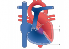
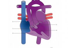
Mesocardia

Mesocardia is that condition in which the longitudinal axis of the heart lies in the mid-sagittal plane and the heart has no apex. Thirteen cases of mesocardia are presented. There were 5 cases of mesoversion, associated with situs solitus of the viscera and normal spleen; 2 cases of presumptive mesoversion, associated with situs inversus of viscera and abnormal spleen; 4 of mixed mesocardia with atrial situs solitus and ventricular inversion, with situs solitus of viscera with or without abnormal spleens; 1 of presumptive mixed mesocardia with atrial situs inversus and normal ventricles, with situs inversus of organs with normal spleen; and 1 case of indeterminate type with situs inversus of viscera and abnormal spleen. Thus, from the stand-point of position of chambers, the type of hearts found in mesocardia resembled those found in dextrocardia and levocardia. If the spleen was normal, then the atrial situs in all cases agreed with that of the viscera and the venous return was normal. Mesocardia reflects that embryonic condition before the formed heart points to the right or left and acquires an apex.
Related Blogs
-
 Jan 1, 1970
Congenital Cystic Adenomatoid Malformation
What is a congenital cystic adenomatoid malformation (CCAM/CPAM)? Congenital cystic adenomatoid malformation (CCAM) is a benign lung lesion that appears before birth as a cyst or mass in the chest...
-
Jan 1, 1970
Tronco arterial comum ou…
Cardiopatia congênita que se caracteriza como o próprio nome diz pela formação de um tronco único saindo do coração. O normal do coração é termos 2 grandes vasos saindo dele,..
-
Jan 1, 1970
Doença de Kawasaki
Na doença de Kawasaki temos uma espécie de inflamação nos pequenos e médios vasos do corpo da criança. Foi descrita pela primeira vez no Japão, em 1962, pelo Dr. Tomisaku..
-
-
Jan 1, 1970
Introducción cardiopatías congénitas
Dr. Mateo Ríos: Médico Pediatra. Cardiólogo Pediatra. Posgrado en Ecocardiografía fetal. Magister en electrofisiología. Actualmente trabaja en el Instituto de Cardiología Integral (Montevideo, Uruguay) en las siguientes..
-
Jan 1, 1970
Circulación fetal, transicional y…
Dr. Mateo Ríos: Médico Pediatra. Cardiólogo Pediatra. Posgrado en Ecocardiografía fetal. Magister en electrofisiología. Actualmente trabaja en el Instituto de Cardiología Integral (Montevideo, Uruguay) en las siguientes..
-
Jan 1, 1970
Embriogénesis Cardíaca
Dr. Mateo Ríos: Médico Pediatra. Cardiólogo Pediatra. Posgrado en Ecocardiografía fetal. Magister en electrofisiología. Actualmente trabaja en el Instituto de Cardiología Integral (Montevideo, Uruguay) en las siguientes..
-
Jan 1, 1970
Fisiopatología de las Cardiopatías…
Dr. Mateo Ríos: Médico Pediatra. Cardiólogo Pediatra. Posgrado en Ecocardiografía fetal. Magister en electrofisiología. Actualmente trabaja en el Instituto de Cardiología Integral (Montevideo, Uruguay) en las siguientes..
-
Jan 1, 1970
Semiología CV
Dr. Mateo Ríos: Médico Pediatra. Cardiólogo Pediatra. Posgrado en Ecocardiografía fetal. Magister en electrofisiología. Actualmente trabaja en el Instituto de Cardiología Integral (Montevideo, Uruguay) en las siguientes..
-
Jan 1, 1970
Clasificación de las Cardiopatías…
Dr. Mateo Ríos: Médico Pediatra. Cardiólogo Pediatra. Posgrado en Ecocardiografía fetal. Magister en electrofisiología. Actualmente trabaja en el Instituto de Cardiología Integral (Montevideo, Uruguay) en las siguientes..
-
Jan 1, 1970
Simple D-Transposition of the…
A heart in which the two main arteries carrying blood away from the heart are reversed. A normal blood pattern carries blood in a cycle: body-heart-lungs-heart-body. When a d-transposition occurs,..
-
Jan 1, 1970
Tetralogy of Fallot (TOF)
Tetralogy of Fallot (TOF) is a cardiac anomaly that refers to a combination of four related heart defects that commonly occur together. The four defects are: − a hole between the right..
-
Jan 1, 1970
Total anomalous pulmonary venous…
Total anomalous pulmonary venous return (TAPVR) is a rare congenital malformation in which all four pulmonary veins do not connect normally to the left atrium. Instead the four pulmonary veins..
-
-
-
Jan 1, 1970
Tetralogy of Fallot (TOF)…
with is a more severe form of TOF, a type of heart defect. It’s a congenital condition, which means it’s something a baby is born with. Babies who have TOF with..
-
-
-
Jan 1, 1970
Superior sinus venosus defect…
The most common venous abnormality of the thorax is persistent left superior vena cava (PLSVC), incidence being less than 0.5%. However, with congenital heart disease, it is about 6.1%. When..
-
Jan 1, 1970
Superior sinus venosus defect…
The most common venous abnormality of the thorax is persistent left superior vena cava (PLSVC), incidence being less than 0.5%. However, with congenital heart disease, it is about 6.1%. When..
-
Jan 1, 1970
Tricuspid atresia with transposed…
Tricuspid atresia is a birth defect of the tricuspid valve, which is the valve that controls blood flow from the right atrium (upper right chamber of the heart) to the..
-
Jan 1, 1970
Tricuspid atresia with patent…
The normal pumping chambers of the heart (the ventricles) must have an inflow valve to let blood in, a good-sized pumping chamber and an outflow to allow blood to exit the..
-
Jan 1, 1970
Tricuspid atresia with congenitally…
Tricuspid atresia is a birth defect of the tricuspid valve, which is the valve that controls blood flow from the right atrium (upper right chamber of the heart) to the..
-
Jan 1, 1970
Tricuspid atresia with atrial…
Tricuspid atresia is a birth defect of the tricuspid valve, which is the valve that controls blood flow from the right atrium (upper right chamber of the heart) to the..
-
Jan 1, 1970
Tricuspid atresia with atrial…
Tricuspid atresia is a birth defect of the tricuspid valve, which is the valve that controls blood flow from the right atrium (upper right chamber of the heart) to the..
-
Jan 1, 1970
Unbalanced complete atrioventricular septal…
The term “unbalance” has been used for decades in atrioventricular septal defect (AVSD) to describe a lack of symmetry between left and right sides of the heart. Even though we..
-
Jan 1, 1970
Supravalvar aortic stenosis, focal…
Supravalvular aortic stenosis(SVAS) is a congenital heart defect. As the name suggests, SVAS means the section of the aorta located just above the aortic valve is narrowed. SVAS accounts for..
-
Jan 1, 1970
Supravalvar aortic stenosis, tubular…
Supravalvular aortic stenosis(SVAS) is a congenital heart defect. As the name suggests, SVAS means the section of the aorta located just above the aortic valve is narrowed. SVAS accounts for..
-
Jan 1, 1970
Subvalvular aortic stenosis, muscular…
Subvalvular aortic stenosis (SAS) is the second most common type of aortic stenosis, accounting for 14% of left ventricular outflow tract (LVOT) obstruction, with valvular aortic stenosis being the most..
-
Jan 1, 1970
Subvalvular aortic stenosis, membranous…
Subvalvular aortic stenosis (SAS) is the second most common type of aortic stenosis, accounting for 14% of left ventricular outflow tract (LVOT) obstruction, with valvular aortic stenosis being the most..
-
Jan 1, 1970
Truncus communis arteriosus Type…
In patients with a TAC only one vessel originates from the heart chambers. Generally the common vessel (truncus) overrides a ventricular septal defect. Depending on the origin of the pulmonary..
-
Jan 1, 1970
Truncus communis arteriosus Type…
In patients with a TAC only one vessel originates from the heart chambers. Generally the common vessel (truncus) overrides a ventricular septal defect. Depending on the origin of the pulmonary..
-
Jan 1, 1970
Truncus communis arteriosus Type…
In patients with a TAC only one vessel originates from the heart chambers. Generally the common vessel (truncus) overrides a ventricular septal defect. Depending on the origin of the pulmonary..
-
-
Jan 1, 1970
Situs inversus
Situs inversus (also called situs transversus or oppositus) is a in which the major are reversed or from their normal positions. The normal arrangement of internal organs is known as . Although cardiac problems are more common, many..
-
Jan 1, 1970
Sinus valsalva aneurysma
Sinus of Valsalva aneurysm (SOVA) is an abnormal dilatation of the aortic root located between the aortic valve annulus and the sinotubular junction. This occurs as a consequence of the..
-
Jan 1, 1970
Single right coronary ostium…
Single coronary artery is a rare congenital anomaly with a quoted incidence of 0.024% to 0.06% (), and may be associated with sudden cardiac death after exercise and angina. We..
-
Jan 1, 1970
Single right coronary ostium…
Single coronary artery is a rare congenital anomaly with a quoted incidence of 0.024% to 0.06% (), and may be associated with sudden cardiac death after exercise and angina. We..
-
Jan 1, 1970
Single right coronary ostium…
Single coronary artery is a rare congenital anomaly with a quoted incidence of 0.024% to 0.06% (), and may be associated with sudden cardiac death after exercise and angina. We..
-
Jan 1, 1970
Single right coronary ostium…
Single coronary artery is a rare congenital anomaly with a quoted incidence of 0.024% to 0.06% (), and may be associated with sudden cardiac death after exercise and angina. We..
-
Jan 1, 1970
Single right coronary ostium…
Single coronary artery is a rare congenital anomaly with a quoted incidence of 0.024% to 0.06% (), and may be associated with sudden cardiac death after exercise and angina. We..
-
Jan 1, 1970
Single coronary artery from…
Single coronary artery is a rare congenital anomaly with a quoted incidence of 0.024% to 0.06% (), and may be associated with sudden cardiac death after exercise and angina. We..
-
Jan 1, 1970
Single left coronary ostium…
Single coronary artery is a rare congenital anomaly with a quoted incidence of 0.024% to 0.06% (), and may be associated with sudden cardiac death after exercise and angina. We..
-
Jan 1, 1970
Simple D-Transposition of the…
Transposition is the condition in which the great arteries take origin from the ventricles in reverse position across (“trans”) the ventricular septum: the aorta from the right ventricle and the..
-
Jan 1, 1970
Ruptured sinus valsalva aneurysma
A sinus of valsalva aneurysm (SOVA) is an abnormal dilatation of the aortic root located between the aortic valve annulus and the sinotubular junction. It occurs as a consequence of..
-
Jan 1, 1970
Ruptured sinus valsalva aneurysm…
Aneurysms of the sinus of Valsalva are extremely rare. Ruptured aneurysms of the sinus of Valsalva are frequently associated with other congenital defects, particularly with ventricular septal defect, aortic valve..
-
Jan 1, 1970
Right ventricular outflow tract…
Congenital heart diseases that cause obstruction of the right ventricular outflow tract are often difficult to diagnose. We report the case of a 49-year-old man who presented with long-standing shortness..
-
Jan 1, 1970
Right ventricular outflow tract…
Congenital heart diseases that cause obstruction of the right ventricular outflow tract are often difficult to diagnose. We report the case of a 49-year-old man who presented with long-standing shortness..
-
-
Jan 1, 1970
Right atrial isomerism, partial…
Partial anomalous pulmonary venous connection (PAPVC) is a rare congenital cardiac defect. As the name suggests, in PAPVC, the blood flow from a few of the pulmonary veins return to..
-
Jan 1, 1970
Right atrial isomerism with…
Background There is a high mortality in infants with right atrial isomerism (RAI). However, less is known about outcome in older children with RAI. This study sought to evaluate those..
-
Jan 1, 1970
Right atrial isomerism with…
The term “atrioventricular septal defect” (AVSD) covers a spectrum of congenital heart malformations characterised by a common atrioventricular junction coexisting with deficient atrioventricular septation. In ostium primum atrial septal defect..
-
Jan 1, 1970
Right pulmonary artery (RPA)…
Pulmonary artery stenosis is a heart defect that causes a narrowing of the pulmonary artery, the large blood vessel that takes blood from the right ventricle of the heart to..
-
-
Jan 1, 1970
Tricuspid valve stenosis
Very little is known about tricuspid valve when compared to other valves, and tricuspid stenosis is rarely described. It most often co-exists with mitral valve pathology especially in patients with..
-
Jan 1, 1970
Uhl anomaly
Uhl's Anomaly is a rare cardiac condition in which there is total absence of right ventricular myocardium resulting in apposition of the endocardium and epicardium. We report a case of..
-
Jan 1, 1970
Turner syndrome
Turner syndrome, a condition that affects only females, results when one of the X chromosomes (sex chromosomes) is missing or partially missing. Turner syndrome can cause a variety of medical..
-
Jan 1, 1970
Single ventricle
A single ventricle is an uncommon occurrence in embryogenesis, which results in the anatomical or functional loss of a ventricular cavity. These malformations are life-threatening and necessitate quick revision to..
-
Jan 1, 1970
Shone Syndrome
Shone syndrome is a collection of eight left-sided obstructive heart lesions. These affect blood flow to and from the left ventricle, or lower left heart chamber. Shone syndrome was identified..
-
Jan 1, 1970
Right cervical arch
Cervical aortic arches are a rare characterized by an elongated, high-lying aortic arch extending at or above the level of the medial ends of the .
-
Jan 1, 1970
Right aortic arch
Right aortic arch anomalies occur in approximately 0.01 to 0.1 percent of the general population. In general, these anomalies do not directly cause any cardiovascular problems. However, in some patients,..
-
Jan 1, 1970
Retroesophagial aortic arch
Right aortic arch is an uncommon anatomic anomaly, which occurs in approximately 0.05% to 0.1% of the population. “Mirror-image” right aortic arch is by itself generally asymptomatic, but it is usually..
-
-
Jan 1, 1970
Pulmonary atresia with ventricular…
The surgical management of pulmonary atresia with ventricular septal defect (VSD), extreme hypoplasia of the pulmonary arteries (PA), major aorto–pulmonary collaterals (MAPCAS) represents a major challenge. Two main basic concepts..
-
-
-
-
Jan 1, 1970
Pulmonary valve stenosis
Pulmonary valve stenosis is a condition in which a deformity on or near your pulmonary valve narrows the pulmonary valve opening and slows the blood flow. The pulmonary valve is..
-
Jan 1, 1970
Persistent left superior caval…
Persistent left superior vena cava (PLSVC) is the most common congenital malformation of thoracic venous return and is present in 0.3 to 0.5% of individuals in the general population. This..
-
Jan 1, 1970
Persistent left superior caval…
Persistent left superior vena cava (PLSVC) is the most common congenital malformation of thoracic venous return and is present in 0.3 to 0.5% of individuals in the general population. This..
-
-
Jan 1, 1970
Patent ductus arteriosus (PDA)
The ductus arteriosus is a normal fetal artery connecting the main body artery (aorta) and the main lung artery (pulmonary artery). The ductus allows blood to detour away from the..
-
Jan 1, 1970
Partial cor triatriatum sinister
Cor Triatriatum Sinister is a rare congenital abnormality, usually diagnosed in childhood; few cases remain asymptomatic and are diagnosed in adulthood. In this review article we focus on classification and..
-
Jan 1, 1970
Partial cor triatriatum sinister…
Cor Triatriatum Sinister is a rare congenital abnormality, usually diagnosed in childhood; few cases remain asymptomatic and are diagnosed in adulthood. In this review article we focus on classification and..
-
Jan 1, 1970
Partial cor triatriatum sinister…
Cor Triatriatum Sinister is a rare congenital abnormality, usually diagnosed in childhood; few cases remain asymptomatic and are diagnosed in adulthood. In this review article we focus on classification and..
-
Jan 1, 1970
Partial atrioventricular septal defect…
[gallery ids="15187,15193,15190"] Atrioventricular (AV) septal defect consists of an ostium primum type atrial septal defect and a common AV valve, with or without an associated inlet (AV septal type) ventricular..
-
Jan 1, 1970
Noonan syndrome
Most people with Noonan syndrome have some form of critical congenital heart disease. The most common heart defect in these individuals is a narrowing of the valve that controls blood flow..
-
Jan 1, 1970
Mitral valve prolapse (Barlow…
Mitral valve prolapse, also called MVP, is a condition in which the two valve flaps of the mitral valve don't close smoothly or evenly, but bulge (prolapse) upward into the left atrium. Mitral valve prolapse is..
-
Jan 1, 1970
Mitral atresia, aortic atresia,…
Although the first-stage Norwood procedure mostly has been used for hypoplastic left heart syndrome, there are other anomalies in which the Norwood procedure can be applied. Since 1991, 18 newborns..
-
Jan 1, 1970
Mitral atresia and hypoplastic…
Mitral atresia is a rare congenital heart defect. This rarity has been reported when compared to atresia of other valves.[] Embryologically, this defect has been attributed to a malaligned ventricular..
-
Jan 1, 1970
Mesoposition
It refers to the degree that the cardiac apex points to the left or right. Position describes the translational relationship or overall location of the heart in the chest. It indicates the hemithorax that the heart predominantly occupies...
-
Jan 1, 1970
Marfan syndrome
Marfan syndrome is a genetic disorder that affects the body’s connective tissue. Connective tissue holds all the body’s cells, organs and tissue together. It also plays an important role in..
-
Jan 1, 1970
Levoposition
These terms purely describe the anatomic position of the left ventricular apex in the chest and their use does not indicate anything about the structure of the heart or the ...
-
Jan 1, 1970
Leopard syndrome
LEOPARD syndrome (LS, OMIM 151100) is a rare multiple congenital anomalies condition, mainly characterized by skin, facial and cardiac anomalies. LEOPARD is an acronym for the major features of this..
-
Jan 1, 1970
Left pulmonary artery (LPA)…
Pulmonary artery stenosis is a narrowing (stenosis) that occurs in the pulmonary artery, a large artery that sends oxygen-poor blood into the lungs to be enriched with oxygen. The narrowing..
-
Jan 1, 1970
Left cervical arch
Cervical aortic arch anomaly is a rare congenital entity. The aortic arch extends into the soft tissues of the neck before turning downward on itself to become the descending aorta...
-
-
-
-
-
-
Jan 1, 1970
Interrupted aortic arch Type…
A rare type of congenital heart disease is an interrupted aortic arch (IAA), which affects approximately 1.5% of congenital heart disease patients. Interrupted aortic arch is an anomaly that can..
-
Jan 1, 1970
Interrupted aortic arch Type…
A rare type of congenital heart disease is an interrupted aortic arch (IAA), which affects approximately 1.5% of congenital heart disease patients. Interrupted aortic arch is an anomaly that can..
-
Jan 1, 1970
Interrupted aortic arch Type…
A rare type of congenital heart disease is an interrupted aortic arch (IAA), which affects approximately 1.5% of congenital heart disease patients. Interrupted aortic arch is an anomaly that can..
-
Jan 1, 1970
Interrupted aortic arch Type…
A rare type of congenital heart disease is an interrupted aortic arch (IAA), which affects approximately 1.5% of congenital heart disease patients. Interrupted aortic arch is an anomaly that can..
-
Jan 1, 1970
Interrupted aortic arch Type…
A rare type of congenital heart disease is an interrupted aortic arch (IAA), which affects approximately 1.5% of congenital heart disease patients. Interrupted aortic arch is an anomaly that can..
-
Jan 1, 1970
Interrupted aortic arch Type…
Interrupted (IAA) is an extremely rare CHD defined as the loss of luminal continuity between the . Its clinical presentation, including , , or severe CHF in the first 2 weeks of life, is..
-
-
Jan 1, 1970
Eisenmenger syndrome with truncus…
Common arterial trunk (CAT), or truncus arteriosus, is a rare form of cyanotic congenital heart disease and is highly associated with DiGeorge syndrome (microdeletion 22q11.2). Prenatal diagnosis is highly feasible,..
-
Jan 1, 1970
Eisenmenger syndrome with single…
Eisenmenger syndrome is the most severe form of pulmonary arterial hypertension and arises on the basis of congenital heart disease with a systemic-to-pulmonary shunt. Due to the chronic slow progressive..
-
Jan 1, 1970
Eisenmenger syndrome with patent…
Development of Eisenmenger syndrome in a known patient of patent ductus arteriosus (PDA) is easy by carefully looking for differential cyanosis and clubbing in upper and lower limbs. It is..
-
Jan 1, 1970
Eisenmenger syndrome with atrioventricular…
Eisenmenger syndrome (ES) is a constellation of symptoms that arise from a congenital heart defect and result in large anatomic shunts. Due to anatomic variations present at birth, hemodynamic forces..
-
Jan 1, 1970
Eisenmenger syndrome with atrial…
Atrial septal defect (ASD) may be rarely associated with Eisenmenger syndrome (ES), the most advanced form of pulmonary vascular disease to complicate a congenital heart disease. In spite of availability..
-
Jan 1, 1970
Eisenmenger syndrome with aorto-pulmonary…
An aortopulmonary (AP) window is a rare cause of Eisenmenger syndrome and results from an abnormal septation of the truncus arteriosus [1]. Most such defects present with early onset congestive heart failure during infancy and adult..
-
Jan 1, 1970
Ebstein anomaly with ventricular…
The Ebstein's anomaly is a malformation of the tricuspid valve, in which the septal and posterior leaflets are attached to the wall of the right ventricle. The usual association is with an atrial septal defect, followed..
-
Jan 1, 1970
Ebstein anomaly with atrial…
A malformed heart valve that does not properly close to keep the blood flow moving in the right direction. Blood may leak back from the lower to upper chambers on..
-
Jan 1, 1970
Ebstein anomaly
Ebstein’s anomaly is a rare congenital heart disorder occurring in ≈1 per 200 000 live births and accounting for <1% of all cases of congenital heart disease. This anomaly was described..
-
Jan 1, 1970
Dysplastic mitral valve with…
Mitral valve dysplasia syndrome is a unique form of congenital heart disease with severe aortic stenosis but normal or enlarged left ventricle secondary to primary mitral valve disease. Increased left..
-
Jan 1, 1970
D-Transposition of the great…
Transposition of the great arteries (TGA) is a pediatric cardiac congenital defect arising from an embryological discordance between the aorta and pulmonary trunk. During cardiac development, the conotruncal septum spirals..
-
Jan 1, 1970
D-Transposition of the great…
Taussig-Bing anomaly is a rare congenital heart malformation that was first described in 1949 by Helen B. Taussig (1898–1986) and Richard J. Bing (1909–). Although substantial improvement has since been..
-
-
Jan 1, 1970
Double outlet right ventricle…
Double outlet right ventricle (DORV) is a heterogeneous group of abnormal ventriculoarterial connections where, by definition, both great arteries (pulmonary artery and aorta) arise primarily from the morphologically right ventricle...
-
Jan 1, 1970
Double outlet right ventricle…
Double-outlet right ventricle (DORV) and subaortic ventricular septal defect (VSD) is defined anatomically as a defect where the entire pulmonary trunk and at least half of the aorta arises from..
-
Jan 1, 1970
Double outlet right ventricle…
The clinical, hemodynamic, angiocardiographic and pathologic findings are presented in an infrequent but surgically correctable type of double outlet right ventricle. This study is based on six cases, one with..
-
Jan 1, 1970
Double outlet right ventricle…
Since Kirklin described the first successful correction of double-outlet right ventricle (DORV), surgical repair has been extended to more complex forms of the malformation . In 1972, Lev and Bharati introduced..
-
Jan 1, 1970
Double outlet left ventricle…
Double-outlet left ventricle (DOLV) is a rare congenital cardiac malformation in which both great arteries originate entirely or predominantly from the morphologic left ventricle. DOLV occurs most commonly in the..
-
Jan 1, 1970
Double inlet right ventricle…
Double outlet right ventricle (DORV) is a rare congenital heart defect, meaning it’s a condition a baby is born with. In DORV, the pulmonary artery and the aorta — the..
-
Jan 1, 1970
Double inlet right ventricle…
Double outlet right ventricle (DORV) is a . There is a malformation of the fetus heart in the womb leading to the right ventricle that has the two major arteries, namely..
-
Jan 1, 1970
Double inlet left ventricle…
Double inlet left ventricle (DILV) is a heart defect that is present from birth (congenital). It affects the valves and chambers of the heart. Babies born with this condition have..
-
-
-
Jan 1, 1970
Double inlet left ventricle…
Double inlet left ventricle (DILV) is a form of functionally univentricular heart where both the left and the right atrium are connected to the morphologically left ventricle. Usually, the morphologically..
-
Jan 1, 1970
Doubled chambered right ventricle
Double-chambered right ventricle (DCRV) was first described in 1858 by TB Peacock, but it is now understood to be a form of congenital heart disease wherein there is a mid-cavitary..
-
Jan 1, 1970
Double aortic arch
Introduction Double aortic arch is the most common type of vascular ring malformation. It involves the complete encirclement and compression of the trachea and/or esophagus by the aortic arch, its..
-
Jan 1, 1970
Discordant criss cross AV…
A heart with a criss-cross atrioventricular (AV) connection is a cardiopathy in which the ventricles are positioned contralaterally to the atria to which they are connected; the ventricular inflow tracts..
-
Jan 1, 1970
Discordant criss cross AV…
A heart with a criss-cross atrioventricular (AV) connection is a cardiopathy in which the ventricles are positioned contralaterally to the atria to which they are connected; the ventricular inflow tracts..
-
Jan 1, 1970
Discordant criss cross AV…
A heart with a criss-cross atrioventricular (AV) connection is a cardiopathy in which the ventricles are positioned contralaterally to the atria to which they are connected; the ventricular inflow tracts..
-
Jan 1, 1970
Discordant criss cross AV…
A heart with a criss-cross atrioventricular (AV) connection is a cardiopathy in which the ventricles are positioned contralaterally to the atria to which they are connected; the ventricular inflow tracts..
-
Jan 1, 1970
Dextroposition
Dextroposition of the : The heart is displaced to the right (from its usual location in the left chest). There is no anatomic alteration in the heart itself, just in its location...
-
Jan 1, 1970
Dextrocardia
Dextrocardia is a rare congenital disorder in which the heart resides on the right side of the thoracic cavity. It is often associated with other development anomalies and, in most..
-
Jan 1, 1970
Unroofed coronary sinus
Unroofed coronary sinus syndrome (URCS) is a rare cardiac anomaly in which a communication occurs between the coronary sinus and the left atrium as a result of the partial or..
-
Jan 1, 1970
Coronary artery fistula congenital
Coronary artery fistulas (CAFs) are abnormal communications of coronary arteries whereby venous circuits bypass the normal capillaries within the myocardium. CAFs are rare, and most affected patients are asymptomatic. However,..
-
Jan 1, 1970
Congenitally corrected transposition of…
Congenitally corrected transposition of the great arteries (CCTGA) is a rare heart defect in which the heart’s lower half is reversed. It is also called L-TGA. It is different from..
-
Jan 1, 1970
Congenitally corrected transposition of…
Congenitally corrected transposition of the great arteries (CCTGA) is a rare congenital heart lesion with varied morphological presentation and can often by asymptomatic. A failing systemic right ventricle (RV) or..
-
Jan 1, 1970
Congenitally corrected transposition of…
Congenitally corrected transposition of the great arteries (CCTGA) is a rare congenital heart lesion with varied morphological presentation and can often by asymptomatic. A failing systemic right ventricle (RV) or..
-
Jan 1, 1970
Congenitally corrected transposition of…
Congenitally corrected transposition of the great arteries (CCTGA) is a rare congenital cardiac anomaly defined by atrio-ventricular and ventriculo-arterial discordance. This malformation makes up less than 1% of congenital heart defects...
-
Jan 1, 1970
Congenitally corrected transposition of…
Congenitally corrected transposition of the great arteries (CCTGA) is a rare congenital cardiac anomaly defined by atrio-ventricular and ventriculo-arterial discordance. This malformation makes up less than 1% of congenital heart defects...
-
Jan 1, 1970
Congenitally corrected transposition of…
Congenitally corrected transposition of the great arteries (CCTGA) is a rare congenital cardiac anomaly defined by atrio-ventricular and ventriculo-arterial discordance. This malformation makes up less than 1% of congenital heart defects...
-
Jan 1, 1970
Congenitally corrected transposition of…
Congenitally corrected transposition of the great arteries (CCTGA) is a rare congenital cardiac anomaly defined by atrio-ventricular and ventriculo-arterial discordance. This malformation makes up less than 1% of congenital heart defects...
-
Jan 1, 1970
Congenitally corrected transposition of…
Congenitally corrected transposition of the great arteries (CCTGA) is a rare complex cardiac anomaly with a wide range of morphologic characteristics. The main underlying disorder is atrioventricular and ventriculoarterial discordance...
-
Jan 1, 1970
Congenitally corrected transposition of…
Congenitally corrected transposition of the great arteries (CCTGA) is a rare congenital cardiac anomaly defined by atrio-ventricular and ventriculo-arterial discordance. This malformation makes up less than 1% of congenital heart defects...
-
Jan 1, 1970
Concordant criss cross AV…
A heart with a criss-cross atrioventricular (AV) connection is a cardiopathy in which the ventricles are positioned contralaterally to the atria to which they are connected; the ventricular inflow tracts..
-
Jan 1, 1970
Concordant criss cross AV…
Criss-cross heart is an extremely rare anomaly, characterized by an abnormal rotation of the ventricular mass along its major axis. It may be associated with any malformation of the heart..
-
Jan 1, 1970
Concordant criss cross AV…
Criss-cross heart (CCH) is a rare of cardiac rotation resulting in crossing of ventricular inlets and drainage of the into contra-laterally located ventricles. The atrio-ventricular (AV) and ventriculo-arterial (VA) connections can be concordant..
-
Jan 1, 1970
Concordant criss cross AV…
Criss-cross heart (CCH) is a rare of cardiac rotation resulting in crossing of ventricular inlets and drainage of the into contra-laterally located ventricles. The atrio-ventricular (AV) and ventriculo-arterial (VA) connections can be concordant..
-
Jan 1, 1970
Complete atrioventricular septal defect…
An atrioventricular septal defect (AVSD) is a heart defect in which there are holes between the chambers of the right and left sides of the heart, and the valves that..
-
Jan 1, 1970
ccTGA with situs inversus,…
Congenitally corrected transposition of the great arteries (ccTGA) is a rare congenital heart defect. There are different subgroups according to the location of the heart in the thorax, apical position..
-
Jan 1, 1970
ccTGA with situs inversus,…
Congenitally corrected transposition of the great arteries (CCTGA) is a rare cardiac anomaly which is characterized by atrioventricular (AV) and ventriculoarterial discordance (transposition of the great arteries), representing less than..
-
Jan 1, 1970
Aortic coarctation; tubular type
Coarctation of the aorta (CoA) is a relatively common defect that accounts for 5-8% of all congenital heart defects. Coarctation of the aorta may occur as an isolated defect or..
-
Jan 1, 1970
Aberrant left pulmonary artery
Aberrant left pulmonary artery, also known as pulmonary sling, represents an anatomical variant characterized by the left pulmonary artery arising from the right pulmonary artery and passing above the right main bronchus and in between the trachea and..
-
Jan 1, 1970
Aberrant left subclavian artery…
Kommerell's diverticulum is most commonly associated with either an aberrant left subclavian artery from a right-sided aortic arch or an aberrant right subclavian artery from a left-sided aortic arch. We..
-
Jan 1, 1970
Aberrant left subclavian artery
Medical genetics. Aberrant subclavian artery, or aberrant subclavian artery syndrome, is a rare anatomical variant of the origin of the right or left subclavian artery. This abnormality is the most common congenital vascular anomaly of the..
-
Jan 1, 1970
Aberrant right subclavian artery
Aberrant right subclavian artery (ARSA) is a rare anomaly, in which the right subclavian artery arises directly from the aortic arch instead of originating from the brachiocephalic artery. This anomaly should be taken into..
-
Jan 1, 1970
Absent pulmonary valve syndrom
Absent pulmonary valve syndrome (APVS) also known as congenital absence of pulmonary valve or pulmonary valve agenesis is a rare outflow tract anomaly of heart. It is defined as absent..
-
Jan 1, 1970
Anomalous left coronary artery…
Anomalous left coronary artery from the pulmonary artery (ALCAPA) is a congenital (present at birth) heart defect in which the left coronary artery arises abnormally from the pulmonary artery. Normally, the left and right coronary arteries arise from the aorta..
-
Jan 1, 1970
Anomalous right coronary artery…
Anomalous right coronary artery (RCA) from pulmonary artery is a rare congenital coronary anomaly. The prevalence is about 0.002% of general population. Anomaly such as aortopulmonary window, tetralogy of Fallot,..
-
-
Jan 1, 1970
Anomalous right coronary artery…
Anomalous right coronary artery (RCA) from pulmonary artery is a rare congenital coronary anomaly. The prevalence is about 0.002% of general population. Anomaly such as aortopulmonary window, tetralogy of Fallot,..
-
Jan 1, 1970
Aortic atresia and hypoplastic…
The spectrum of hypoplastic left heart syndrome spans a continuum from borderline hypoplasia to extreme forms with retrograde blood flow from the ductus arteriosus to the cerebral and coronary arteries...
-
Jan 1, 1970
Aortic coarctation with biscuspid…
Prevalence of congenital heart disease (CHD) is constantly increasing during the last decades in line with the treatment options for patients ranging from the surgical as well to the interventional..
-
-
Jan 1, 1970
Aortic coarctation; focal type
Coarctation of aorta (CoA) can be simply defined as cardiac abnormality resulting in obstruction to the blood flow in the aorta. CoA can occur at any region in the thoracic..
-
Jan 1, 1970
Aortopulmonary window
Aortopulmonary window (APW) or aortopulmonary septal defect, first described by Elliotson in third decade of last century,[] is a cardiac abnormality that results from abnormal communication between the proximal aorta..
-
Jan 1, 1970
Aortic regurgitation
Abnormal aortic valve development is one of the most threatening congenital heart diseases. Like a thief in the night, the patient might be struck by endocarditis after a visit to..
-
Jan 1, 1970
Congenital aortic stenosis
Aortic stenosis refers to a condition that causes obstruction to blood flow between the left ventricle and the aorta. There are a variety of causes, including muscular obstruction below the..
-
Jan 1, 1970
Bicuspid aortic valve with…
Bicuspid aortic valve (BAV) is the most common congenital cardiac abnormality, affecting approximately 1%-2% of the general population. (1,2) Adverse cardiac outcomes related to the valve and/or root (3) put..
-
Jan 1, 1970
Bicuspid aortic valve with…
Subvalvular aortic stenosis (SAS), also called subaortic stenosis, is a rare disorder seen in infants. In most cases, it involves the presence of a membrane that is typically muscular just..
-
Jan 1, 1970
Bicuspid aortic valve with…
Bicuspid aortic valve (BAV) is the most common congenital cardiac abnormality, occurring in 0.5%-1.4% of the population; this anomaly is sporadically transmitted genetically by an autosomal-dominant pathway, with a 3:1..
-
Jan 1, 1970
Bicuspid aortic valve with…
Bicuspid aortic valve (BAV) and congenital aortic stenosis are two types of heart defects that may be present at birth. They can occur separately or together. In some cases, bicuspid aortic valve causes another condition called aortic valve..
-
Jan 1, 1970
Bicuspid aortic valve with…
Congenital bicuspid aortic valve is a relatively rare malformation. It is reported that the presence of this anomaly predisposes the patient to development of true aortic aneurysms or dissecting aortic..
-
Jan 1, 1970
Bicuspid aortic valve with…
Bicuspid aortic valve (BAV) is the most common congenital heart defect with a prevalence of 1–2% and most commonly BAV is found in males with a rate of 1:2 varying..
-
Jan 1, 1970
Bicuspid aortic valve
The aortic valve allows oxygen-rich blood to flow from the heart to the aorta. It prevents the blood from flowing back from the aorta into the heart when the pumping..
-
Jan 1, 1970
Total anomalous pulmonary venous…
Total anomalous pulmonary venous connection (TAPVC) is a very rare congenital heart disease (CHD), reported in 1-2.6% of all congenital heart disease (-). The onset can be abrupt with cardiopulmonary..
-
-
Jan 1, 1970
Total anomalous pulmonary venous…
Total anomalous pulmonary venous connection (TAPVC) develops when the primordial pulmonary vein fails to unite with the plexus of veins surrounding the lung buds. In 1959, Darling and associates[] proposed..
-
Jan 1, 1970
Total anomalous pulmonary venous…
In this type of TAPVR, the pulmonary veins first come together behind the heart and then drain upwards to an abnormal “vertical vein.” This vertical vein joins the innominate vein which connects to the right superior vena cava and drains to the..
-
Jan 1, 1970
Total anomalous pulmonary venous…
Anomalous pulmonary venous connection (APVC) is an uncommon congenital anomaly in which pulmonary venous blood flows directly into the right side of the heart or into the systemic veins. The..
-
Jan 1, 1970
Scimitar syndrome
Scimitar syndrome consists of a partial anomalous pulmonary venous drainage of right lung, right lung hypoplasia, dextraposition of heart, and anomalous systemic arterial supply from aorta or one of its..
-
Jan 1, 1970
Partial anomalous pulmonary venous…
Partial anomalous pulmonary venous return, sometimes called partial anomalous pulmonary venous connection, is a heart defect present at birth (congenital) in which some of the pulmonary veins carrying blood from..
-
-
Jan 1, 1970
Partial anomalous pulmonary venous…
Partial anomalous pulmonary vein connection (PAPVC) is a rare congenital abnormal cardiac defect involving the pulmonary veins draining into the right atrium (RA) directly or indirectly by venous connection. Nowadays, these patients are diagnosed when still..
-
Jan 1, 1970
Partial anomalous pulmonary venous…
Partial anomalous venous connection (PAPVC) is a rare congenital heart disease where the blood flow from one or more pulmonary veins (but not all) returns to the right atrium or systemic venous circulation and is..
-
Jan 1, 1970
Partial anomalous pulmonary venous…
Partial anomalous pulmonary venous connection (PAPVC) is a rare congenital cardiac defect. As the name suggests, in PAPVC, the blood flow from a few of the pulmonary veins return to..
-
Jan 1, 1970
Partial anomalous pulmonary venous…
Partial anomalous pulmonary venous connection (PAPVC) is a rare congenital cardiac defect. As the name suggests, in PAPVC, the blood flow from a few of the pulmonary veins return to the right atrium instead..
-
Jan 1, 1970
Mitral valve prolapse (Barlow…
Barlow’s syndrome has become a regular, often-used and very often misused diagnosis. Its description followed extensive, prolonged and detailed clinical observation by JB Barlow and his co-workers. This major research..
-
Jan 1, 1970
Partial anomalous pulmonary venous…
Partial anomalous venous connection (PAPVC) is a rare congenital heart disease where the blood flow from one or more pulmonary veins (but not all) returns to the right atrium or..
-
Jan 1, 1970
Mitral valve prolapse
Mitral valve prolapse, also called MVP, is a condition in which the two valve flaps of the mitral valve don't close smoothly or evenly, but bulge (prolapse) upward into the..
-
-
Jan 1, 1970
”ECMO, El éxito de…
ECMO, El éxito de una terapia MÓDULO II – “ECMO, state of the art” MODERADOR Principal: Dr. Gregorio Rábago MD. Servicio Cirugía Cardiaca C.U.N., Pamplona/Madrid MODERADOR Chat: Dr. José Lopez...
-
-
-
Jan 1, 1970
Cor triatriatum sinister with…
Cor triatriatum sinister is a relatively rare congenital condition in which the left atrium is bisected by a fibromuscular membrane into two distinct chambers. There are multiple hypotheses for the..
-
Jan 1, 1970
Cor triatriatum sinister with…
Cor Triatriatum Sinister is a rare congenital abnormality, usually diagnosed in childhood; few cases remain asymptomatic and are diagnosed in adulthood. In this review article we focus on classification and..
-
Jan 1, 1970
Cor triatriatum sinister
The cor triatriatum sinister is a rare congenital heart defect, the product of an embryonic defect in the incorporation of the common pulmonary vein to the left atrium, this being..
-
Jan 1, 1970
Supravalvular mitral ring with…
Supravalvar mitral ring is a circumferential ridge or membrane arising from the left atrial wall overlying the mitral valve and frequently attached to the mitral valve. Variable in thickness and extent,..
-
Jan 1, 1970
Inferior sinus venosus defect…
Background Sinus venosus defect (SVD) is an unusual type of interatrial communication (IAC) and is virtually always associated with partial anomalous pulmonary venous drainage (PAPVD) of the right pulmonary veins..
-
Jan 1, 1970
Muscular ventricular septal defect…
A ventricular septal defect (VSD) is a communication between the interventricular chambers. Muscular ventricular septal defects have exclusively muscular borders and are more likely to be multiple. Their location and..
-
Jan 1, 1970
Muscular ventricular septal defect…
A ventricular septal defect (VSD) is an opening in the interventricular septum, causing a shunt between ventricles. Large defects result in a significant left-to-right shunt and cause dyspnea with feeding..
-
Jan 1, 1970
Perimembranous ventricular septal defect…
Perimembranous ventricular septal defects (VSDs) are located in the left ventricle outflow tract beneath the aortic valve. They are the most common subtype in the United States, occurring in 75-80% of..
-
Jan 1, 1970
Perimembranous ventricular septal defect…
Perimembranous ventricular septal defects (VSDs) are located in the left ventricle outflow tract beneath the aortic valve. They are the most common VSD subtype in the United States, occurring in 75-80% of cases. Defects may extend into..
-
Jan 1, 1970
Doubly committed ventricular septal…
Doubly committed subarterial ventricular septal defect (VSD) is a unique type of VSD, located beneath the aortic and pulmonary valves, accounting for about 5–7% of all VSD autopsy findings. It is also called supracristal or infundibular VSD..
-
Jan 1, 1970
Doubly committed ventricular septal…
Background: The morphology of ventricular septal defects (VSDs) that are doubly committed and juxtaarterial places the patient at risk for aortic valvar prolapse and aortic valvar insufficiency (AI). Surgical repair of..
-
Jan 1, 1970
Complete atrioventricular septal defect…
Atrioventricular septal defects (AVSD) are a relatively common family of congenital heart defects. Also known as atrioventricular canal defects or endocardial cushion defects, they account for about 5 percent of..
-
Jan 1, 1970
Complete atrioventricular septal defect…
Female infant; born on term with 2.6 kg, normal delivery. Mother; fourth pregnancy, denied the use of medication during pregnancy. Presented respiratory discomfort at birth, evolving with neonatal infection and..
-
Jan 1, 1970
Unbalanced complete atrioventricular septal…
Background: Management of right-dominant atrioventricular septal defect (AVSD) remains a challenge given the spectrum of ventricular hypoplasia. The purpose of this study was to assess whether reported echocardiographic indices and additional..
-
Jan 1, 1970
Unbalanced complete atrioventricular septal…
Unbalance in atrioventricular septal defect can be found in more than one anatomic level and in different degrees at each level. The definition of “unbalance” has historically been focused in..
-
Jan 1, 1970
Complete atrioventricular septal defect…
Repair for tetralogy of Fallot (TOF) with complete atrioventricular septal defect (CAVSD) has been reported with good early and intermediate outcomes. Morbidity, however, remains significantly high. To date, repair of..
-
Jan 1, 1970
Partial atrioventricular septal defect…
Atrioventricular (AV) septal defect consists of an ostium primum type atrial septal defect and a common AV valve, with or without an associated inlet (AV septal type) ventricular septal defect..
-
Jan 1, 1970
Dysplastic mitral valve with…
syndrome is a unique form of left-sided heart disease characterized by aortic outflow , dilated , dysplastic/incompetent mitral valve, and a restrictive/intact . Patients with this constellation of abnormalities have been managed in..
-
Jan 1, 1970
Parachute mitral valve with…
Congenital (MS) results from a variety of anatomic anomalies in the pediatric population, and it is commonly associated with parachute (PMV),, an abnormality in which the mitral valve (MV) chordae insert into a..
-
Jan 1, 1970
Levocardia
In levocardia, the heart is predominantly in the left hemithorax with a leftward apex. In , the heart is predominantly in the right hemithorax. Primary dextrocardia is defined as a condition in..
-
Jan 1, 1970
Inferior sinus venosus defect…
Inferior sinus venosus defects (SVDs) are rare imperfections located in the inferior portion of the atrial septum, leading to an overriding inferior vena cava (IVC) and an interatrial connection. These..
-
Jan 1, 1970
Superior sinus venosus defect…
Partial anomalous venous connection (PAPVC) is a rare congenital heart disease where the blood flow from one or more pulmonary veins (but not all) returns to the right atrium or systemic venous circulation and is..
-
-
Jan 1, 1970
Superior sinus venosus defect…
Surgical repair of sinus venosus defect (SVD) with partial anomalous pulmonary venous connection (PAPVC) to the superior vena cava (SVC) or the right atrium (RA) should avoid long term complications..
-
Jan 1, 1970
Unroofed coronary sinus
Sinus venosus defects (SVDs), originally described in 1858, represent about 2%–10% of all atrial septal defects. The remainder is composed of ostium secundum (70%), ostium primum (20%) and unroofed coronary sinus (<1%)..
-
Jan 1, 1970
Unroofed coronary sinus with…
The unroofed coronary sinus is a spectrum of cardiac anomalies in which part or all of the common wall between the coronary sinus and the left atrium is absent. Most..
-
Jan 1, 1970
Atrial septal defect (ASD)…
What is it? A "hole" in the wall that separates the top two chambers of the heart. This defect allows oxygen-rich blood to leak into the oxygen-poor blood chambers in..
-
Jan 1, 1970
Minimally invasive ASD repair.…
Educational video. Axillary approach has many advantages in congenital heart surgery - cosmetical, physical, psychological benefits for children.
-
-
-
Jan 1, 1970
Situs solitus
Visceroatrial situs refers to the position and configuration of the cardiac atria, the tracheobronchial tree, and the thoracoabdominal viscera. Accurate determination of situs is essential because anomalies of situs are..
-
-
-
-
-
-
-
-
-
-
-
-
-
-
-
-
-
Jan 1, 1970
Tetralogy of fallot with…
Congenital absence of pulmonary valve syndrome (APV) represents a fascinating and unique variant of congenital heart disease. It was Chever in 1847 who first described this unique structural heart defect...
-
Jan 1, 1970
PATENT DUCTUS ARTERIOSUS (PDA)
From week 6 of fetal life until birth, the ductus is responsible for most of the right ventricular outflow. Normally, functional closure of the ductus arteriosus occurs by about 15..
-
Jan 1, 1970
Patent foramen ovale (PFO)
A hole in your heart would seem to be the very definition of a "problem." Yet more than a quarter of the population has one, and for most it causes..