Aortic coarctation; focal type
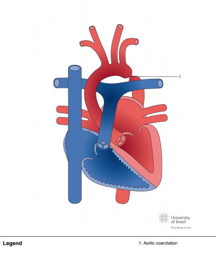
Coarctation of aorta (CoA) can be simply defined as cardiac abnormality resulting in obstruction to the blood flow in the aorta. CoA can occur at any region in the thoracic and abdominal aorta. Most common location for CoA is just distal to the left subclavian artery at the point where ductus arteriosus connects to the aorta. Typically there is presence of medial thickening with “shelf like” tissue protruding in the lumen of aorta from the posterior aortic wall [1, 2]. CoA was first described by Giovanni Morgagni, an Italian anatomist, in the 18th century. First therapeutic surgical intervention was performed for this condition in the year 1944 by Dr. Crafoord [2, 3]. CoA carried a very poor prognosis in the presurgical era with median survival age of mere 31 years [4]. Transcatheter balloon angioplasty was introduced for CoA management in early 1980s by Singer et al. [5]. Later in the decade transcatheter endovascular stent therapy was used for the management of CoA [6]. Now, for about 75 years since first surgical intervention for CoA and significant advances in transcatheter therapy, natural history of this disease has significantly changed. Most of these patients are making it to the adulthood.