Perimembranous ventricular septal defect (VSD)
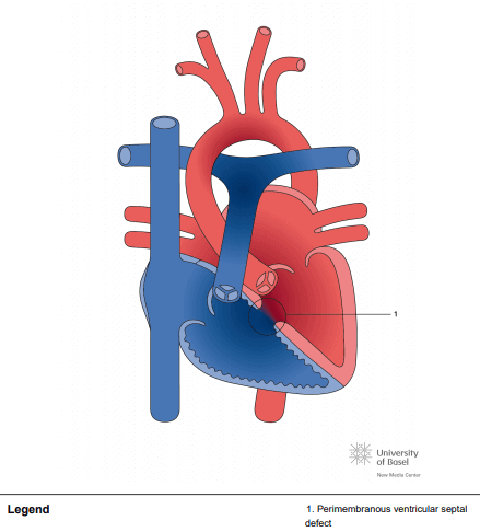
Perimembranous ventricular septal defects (VSDs) are located in the left ventricle outflow tract beneath the aortic valve. They are the most common VSD subtype in the United States, occurring in 75-80% of cases. Defects may extend into adjacent portions of the ventricular septum. When tissue forms on the right ventricular septal surface (often thought to be tricuspid valvular in origin), it is termed an aneurysm of the membranous septum. Such tissue serves as a mechanism of restriction or spontaneous closure. The defect may be partially or completely occluded by the septal leaflet of the tricuspid valve. (See Epidemiology, Prognosis, and Treatment.)
Normal closure of the ventricular septum occurs through multiple concurrent embryologic mechanisms that help to close the septum’s membranous portion: (1) downward growth of the conotruncal ridges forming the outlet septum, (2) growth of the endocardial cushions forming the inlet septum, and (3) growth of the muscular septum forming the apical and midmuscular portions of the septum.