Discover our section specialized in congenital heart disease pathologies, where you can find more than 200 pathologies.
Collaboration with Prof. Daniel Tobler and University of Basel

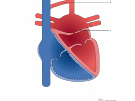

Cor triatriatum sinister is a relatively rare congenital condition in which the left atrium is bisected by a fibromuscular membrane into two distinct chambers. There are multiple hypotheses for the embryologic origin of this congenital defect. The presentation of patients can be during infancy, childhood, or adulthood, and this is..
Read More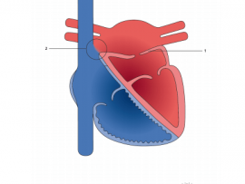
Cor Triatriatum Sinister is a rare congenital abnormality, usually diagnosed in childhood; few cases remain asymptomatic and are diagnosed in adulthood. In this review article we focus on classification and etiologies, clinical manifestations, novel diagnostic modalities and treatment of Cor triatriatum Sinister.
Read More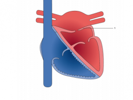
The cor triatriatum sinister is a rare congenital heart defect, the product of an embryonic defect in the incorporation of the common pulmonary vein to the left atrium, this being divided into two separate chambers by a fibromuscular membrane. We report the case of 5 months old boy with recurrent..
Read More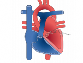
Supravalvar mitral ring is a circumferential ridge or membrane arising from the left atrial wall overlying the mitral valve and frequently attached to the mitral valve. Variable in thickness and extent, the ring ranges from a thin membrane to a thick discrete fibrous ridge. The membranous variety may be difficult to detect..
Read More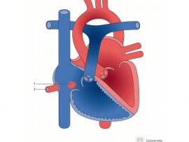
Background Sinus venosus defect (SVD) is an unusual type of interatrial communication (IAC) and is virtually always associated with partial anomalous pulmonary venous drainage (PAPVD) of the right pulmonary veins (RPV) to the superior vena cava (SVC) or right atrium (RA). However, its definite morphogenesis is still elusive, and diagnostic..
Read More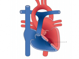
A ventricular septal defect (VSD) is a communication between the interventricular chambers. Muscular ventricular septal defects have exclusively muscular borders and are more likely to be multiple. Their location and multiplicity can sometimes make them a very challenging clinical problem.
Read More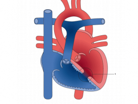
A ventricular septal defect (VSD) is an opening in the interventricular septum, causing a shunt between ventricles. Large defects result in a significant left-to-right shunt and cause dyspnea with feeding and poor growth during infancy. A loud, harsh, holosystolic murmur at the lower left sternal border is common. Recurrent respiratory..
Read More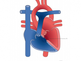
Perimembranous ventricular septal defects (VSDs) are located in the left ventricle outflow tract beneath the aortic valve. They are the most common subtype in the United States, occurring in 75-80% of cases. Defects may extend into adjacent portions of the ventricular septum. When tissue forms on the right ventricular septal surface..
Read More