Summary of “Personalized ventilation guided by electrical impedance tomography with increased PEEP improves ventilation‐perfusion matching in asymmetrical airway closure and contralateral pulmonary embolism during veno‐venous extracorporeal membrane oxygenation: A case report”
Abstract
This case report describes a novel use of electrical impedance tomography (EIT) to guide personalized PEEP adjustments in a 54-year-old patient on ECMO with right lung pneumonia and contralateral pulmonary embolism (PE). By using EIT to assess ventilation and perfusion in real time, clinicians adjusted PEEP to exceed the airway opening pressure (AOP) of the injured lung, improving V/Q matching, oxygenation, and hemodynamics without compromising cardiovascular stability. The findings illustrate the value of individualized, physiology-based ventilation strategies in complex critical care scenarios.

Key Points:
-
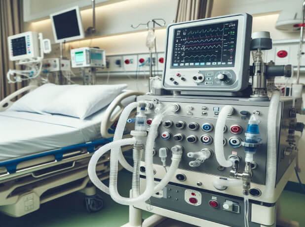
Clinical Background: The patient had severe refractory hypoxemia caused by asymmetrical lung injury—right-sided pneumonia and left-sided PE—complicating ventilator management and requiring VV-ECMO.
-
Initial EIT Findings at PEEP 12 cmH₂O: EIT revealed ventilation was predominantly in the left lung, while perfusion remained primarily in the right lung, creating severe V/Q mismatch due to dead space and shunt physiology.
-
PEEP Personalization Based on AOP: EIT-guided low-flow inflation identified differing AOPs: 8 cmH₂O in the left lung and 16 cmH₂O in the right (pneumonia-affected) lung. PEEP was increased to 20 cmH₂O to maintain right lung recruitment.
-
Impact on Lung Mechanics: Higher PEEP improved EELI (end-expiratory lung impedance), especially in the right lung, indicating more sustained alveolar recruitment. Some overdistension was observed in ventral left lung regions.
-
Ventilation-Perfusion Coupling Improvement: V/Q matching improved from 29% to 41.7%, while unmatched perfusion (shunt) dropped from 21.7% to 12.8%, and unmatched ventilation (dead space) decreased from 49.3% to 45.5%.
-
Enhanced Gas Exchange: PaO₂ improved from 59 to 66 mmHg after PEEP titration, and SvO₂ rose from 84% to 85.5%, despite unchanged ECMO settings, suggesting improved pulmonary oxygenation efficiency.
-
Cardiac Output and Hemodynamics: Cardiac output increased from 8.5 to 9.8 L/min with PEEP 20 cmH₂O, alongside a significant drop in pulmonary vascular resistance (from 169 to 122 dyn•s/cm⁵•m²), suggesting favorable right ventricular unloading.
-
PE Confirmation and Resolution: EIT findings suggested a left-sided PE, which was later confirmed by CT scan. The patient underwent thromboaspiration, leading to further improvements in PaO₂ and FiO₂ weaning.
-
Clinical Course and Outcome: The patient improved steadily, was weaned off ECMO by day 10, extubated by day 17, and discharged in stable condition on day 36. The underlying diagnosis was cryptogenic organizing pneumonia.
-
Clinical Implication of EIT: EIT enabled real-time assessment of differential lung mechanics and perfusion, enabling physiology-based, bedside personalization of PEEP. It also served as a noninvasive early diagnostic clue for PE.

Conclusion
This case highlights the clinical value of bedside EIT in tailoring mechanical ventilation in complex ICU patients. By adjusting PEEP to exceed the AOP of the consolidated lung, clinicians improved oxygenation and V/Q matching without compromising hemodynamics. Moreover, EIT identified perfusion defects consistent with PE, prompting timely imaging and intervention. This illustrates the potential of EIT as both a therapeutic and diagnostic tool in precision critical care.


Watch the following video on “Electrical Impedance Tomography Guided APRV” by Respiratory Associates
Open Access This article is licensed under a Creative Commons Attribution 4.0 International License, which permits use, sharing, adaptation, distribution and reproduction in any medium or format, as long as you give appropriate credit to the original author(s) and the source, provide a link to the Creative Commons licence, and indicate if changes were made. The images or other third party material in this article are included in the article’s Creative Commons licence, unless indicated otherwise in a credit line to the material. If material is not included in the article’s Creative Commons licence and your intended use is not permitted by statutory regulation or exceeds the permitted use, you will need to obtain permission directly from the copyright holder. To view a copy of this licence, visit http://creativecommons.org/licenses/by/4.0/.