How to manage differential hypoxemia during veno-arterial extracorporeal membrane oxygenation
Abstract
Over the past decade, veno-arterial extracorporeal membrane oxygenation (VA-ECMO) has expanded rapidly as a salvage strategy to provide temporary circulatory and respiratory support allowing cardiac function recovery or bridging to additional therapeutic alternatives in patients with refractory cardiogenic shock.
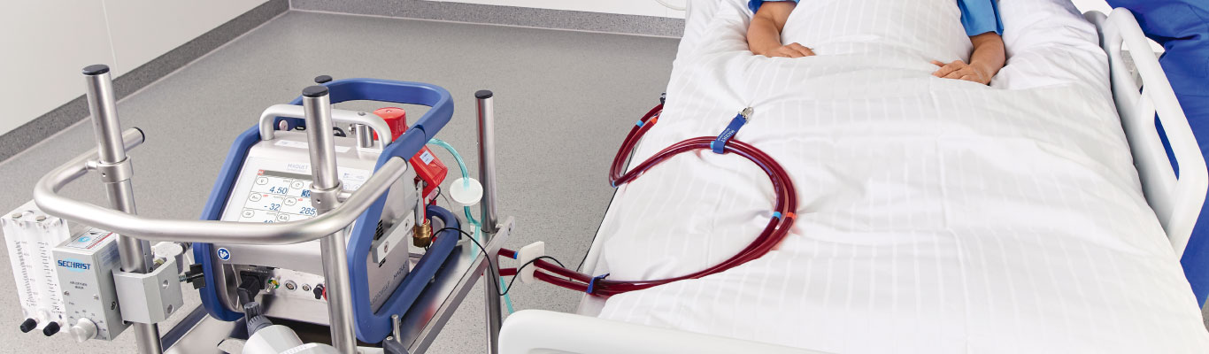
Peripheral access using the femoral vein and artery (FA) is the most widely used method for the initiation of VA-ECMO in adult patients.1 When venous blood is drained from the inferior caval vein (IVC), the patient is at risk of developing differential oxygenation meaning a low oxygenation of the upper part of the body, while the lower part has high oxygenation.
Differential oxygenation might cause insufficient oxygen supply to the heart and brain and affect the prognosis of patients. Understanding mechanism and appropriate management of differential oxygenation are critical to favorable outcomes in patients undergoing peripheral VA-ECMO.
Dual circulation is underrecognized physiological phenomenon and has been proposed as the major mechanism for differential hypoxia in patients with femoral VA-ECMO (IVC-FA). In peripheral VA-ECMO, blood is always returned to the femoral artery, and the retrograde ECMO flow in the descending aorta meets the blood ejected from the left ventricle (LV) in a mixing zone somewhere along the aorta.
The location of the mixing zone depends on strengths of the native heart and extracorporeal pump, and will determine which areas of the body are perfused with blood ejected from the LV and which areas of the body are perfused by the reinfused blood from the ECMO circuit. In patients with extremely low native cardiac output, the mixing zone might be located near the aortic root. When the native cardiac output increases, the mixing zone might be located in the aortic arch or descending aorta. In addition, a change in ECMO flow will bring a corresponding change in mixing zone.
Therefore, physiology is changed from one serial pump to two circuits pumping in parallel. In patients with residual cardiac function and severely impaired pulmonary function, the mixing zone might move to distal aortic arch or descending aorta, and the blood ejected from the LV is poorly oxygenated, predominantly supporting the upper part of the body (heart and brain), drains into superior caval vein (SVC), and is pumped through the sick native lungs to the left atrium and LV without being mixed with the oxygenated blood from ECMO, whereas the hyper-oxygenated blood from ECMO enters the descending aorta, supporting the lower body (splanchnic circulation, legs, etc.) and drains into the IVC and further to the ECMO circuit. This phenomenon has been called “differential oxygenation”.