
Complex Heart-Lung Procurement and Back-Table Reconstruction


Navigating the Anatomical Storm: Complex Heart-Lung Procurement and Back-Table Reconstruction
In the high-stakes arena of cardiothoracic transplantation, procurement surgery is often treated as a standardized exercise. Surgeons rely on predictable anatomy to efficiently protect and harvest donor organs. However, when a donor presents with a cluster of rare, compounding congenital vascular anomalies, the routine procurement transforms into a masterclass in surgical adaptability.
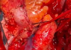
Encountering a donor with an absent brachiocephalic vein, duplicate superior venae cavae (SVC), a bovine aortic arch, and a double aortic arch requires a radical departure from standard protocols. Successfully navigating this anatomical storm demands meticulous in-situ strategy and creative, ex-vivo «back-table» reconstruction to ensure the organs are implantable.
The Anatomical Landscape: A Perfect Storm of Anomalies.
To appreciate the complexity of this procurement, one must visualize how these four distinct anomalies alter the superior mediastinum:
1. Duplicate SVC with Absent Brachiocephalic Vein: Normally, the left and right brachiocephalic veins unite to form a single right-sided SVC. Here, the lack of a cross-communicating brachiocephalic vein means the left upper extremity and head drain via a persistent left SVC (PLSVC), which typically empties into the coronary sinus. The right SVC drains the right side normally.
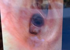
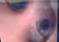
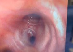
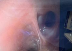
2. Double Aortic Arch: The ascending aorta splits into two arches (forming a vascular ring around the trachea and esophagus) before reuniting into the descending thoracic aorta.
3. Bovine Aorta: Coexisting within this complex, a «bovine» pattern typically implies that the brachiocephalic artery and the left common carotid artery share a common origin, further crowding the arch anatomy.
In-Situ Strategy: Safe Cannulation and Cross-Clamping
The primary objective during in-situ procurement is achieving uniform organ preservation (plegia) without causing mechanical injury or distension to the heart and lungs.
1. Mobilization and Dissection
Standard median sternotomy must be extended superiorly. Wide mobilization of the pericardium and the pleural reflections is mandatory. The surgeon must carefully isolate both the right SVC and the left SVC to ensure adequate control. The double aortic arch must be dissected free from surrounding tissue, paying close attention to the recurrent laryngeal nerves trapped within the vascular ring.
2. Venting and Plegia Delivery
The Venous Challenge: Because there is no brachiocephalic vein connecting the two sides, separate venting strategies or dual-cannulation planning for the recipient must be considered. For standard donor cardioplegia, a single cannula in the ascending aorta suffices, provided it is placed proximal to the bifurcation of the double arch.
The Arterial Challenge: Cross-clamping a double aortic arch is dangerous. Clamping just one arch will leave the other patent, leading to incomplete arrest, warm myocardial perfusion, and inadequate lung preservation via bronchial collaterals.
Encountering a donor with an absent brachiocephalic vein, duplicate superior venae cavae (SVC), a bovine aortic arch, and a double aortic arch requires a radical departure from standard protocols. Successfully navigating this anatomical storm demands meticulous in-situ strategy and creative, ex-vivo «back-table» reconstruction to ensure the organs are implantable.
The Anatomical Landscape: A Perfect Storm of Anomalies.
To appreciate the complexity of this procurement, one must visualize how these four distinct anomalies alter the superior mediastinum:
1. Duplicate SVC with Absent Brachiocephalic Vein: Normally, the left and right brachiocephalic veins unite to form a single right-sided SVC. Here, the lack of a cross-communicating brachiocephalic vein means the left upper extremity and head drain via a persistent left SVC (PLSVC), which typically empties into the coronary sinus. The right SVC drains the right side normally.
2. Double Aortic Arch: The ascending aorta splits into two arches (forming a vascular ring around the trachea and esophagus) before reuniting into the descending thoracic aorta.
3. Bovine Aorta: Coexisting within this complex, a «bovine» pattern typically implies that the brachiocephalic artery and the left common carotid artery share a common origin, further crowding the arch anatomy.
In-Situ Strategy: Safe Cannulation and Cross-Clamping
The primary objective during in-situ procurement is achieving uniform organ preservation (plegia) without causing mechanical injury or distension to the heart and lungs.
1. Mobilization and Dissection
Standard median sternotomy must be extended superiorly. Wide mobilization of the pericardium and the pleural reflections is mandatory. The surgeon must carefully isolate both the right SVC and the left SVC to ensure adequate control. The double aortic arch must be dissected free from surrounding tissue, paying close attention to the recurrent laryngeal nerves trapped within the vascular ring.
2. Venting and Plegia Delivery
The Venous Challenge: Because there is no brachiocephalic vein connecting the two sides, separate venting strategies or dual-cannulation planning for the recipient must be considered. For standard donor cardioplegia, a single cannula in the ascending aorta suffices, provided it is placed proximal to the bifurcation of the double arch.
The Arterial Challenge: Cross-clamping a double aortic arch is dangerous. Clamping just one arch will leave the other patent, leading to incomplete arrest, warm myocardial perfusion, and inadequate lung preservation via bronchial collaterals.
Unifying the Double Aortic Arch & Bovine Origin
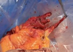
The recipient cannot safely accommodate a double aortic arch. On the back table, the surgeon must surgically alter the donor aorta:
The minor or non-dominant arch (often the left) is typically divided or unroofed.
The remaining dominant arch is opened longitudinally, incorporating the origins of the bovine carotids.
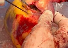
Using a donor pericardial patch or a portion of the divided arch wall, the surgeon reconstructs the aorta into a single, unified, wide-mouth aortic cuff that resembles a normal aortic arch, ready for standard anastomosis.
2. Managing the Dual SVCs
If the heart and lungs are going to the same recipient, the dual SVCs can either be kept intact (if the recipient has similar anatomy) or reconstructed. If the organs are being split (heart to one recipient, lungs to another):
The Heart: The right SVC is kept with a standard cuff. The persistent left SVC (which drains into the coronary sinus) must be carefully dissected. The surgeon must ensure that preserving the PLSVC patch does not compromise the left atrial cuff needed for the lung recipient.
The Lungs: If being utilized for bilateral lung transplantation, the pulmonary veins are divided into standard left and right atrial cuffs.
3. Re-creating the Venous Confluence
Because the donor lacked a brachiocephalic vein, the back-table surgeon can use donor innominate or iliac vein grafts (harvested separately from the donor) to construct a Y-shaped venous conduit. This connects the left and right SVC stumps into a single channel, ensuring that when the recipient is implanted, a single superior vena caval anastomosis can be performed.
Conclusion
Procuring organs from a donor with an absent brachiocephalic vein, dual SVCs, a bovine aorta, and a double arch represents the absolute pinnacle of surgical problem-solving. Success hinges on recognizing the anomalies instantly, modifying the cross-clamp strategy to ensure uniform organ preservation, and utilizing precise ex-vivo back-table reconstruction. By reshaping these congenital anomalies into standard surgical cuffs, transplant surgeons rescue viable organs from the brink of decline, turning an anatomical anomaly into a life-saving triumph.
The recipient cannot safely accommodate a double aortic arch. On the back table, the surgeon must surgically alter the donor aorta:
The minor or non-dominant arch (often the left) is typically divided or unroofed.
The remaining dominant arch is opened longitudinally, incorporating the origins of the bovine carotids.
Using a donor pericardial patch or a portion of the divided arch wall, the surgeon reconstructs the aorta into a single, unified, wide-mouth aortic cuff that resembles a normal aortic arch, ready for standard anastomosis.
2. Managing the Dual SVCs
If the heart and lungs are going to the same recipient, the dual SVCs can either be kept intact (if the recipient has similar anatomy) or reconstructed. If the organs are being split (heart to one recipient, lungs to another):
The Heart: The right SVC is kept with a standard cuff. The persistent left SVC (which drains into the coronary sinus) must be carefully dissected. The surgeon must ensure that preserving the PLSVC patch does not compromise the left atrial cuff needed for the lung recipient.
The Lungs: If being utilized for bilateral lung transplantation, the pulmonary veins are divided into standard left and right atrial cuffs.
3. Re-creating the Venous Confluence
Because the donor lacked a brachiocephalic vein, the back-table surgeon can use donor innominate or iliac vein grafts (harvested separately from the donor) to construct a Y-shaped venous conduit. This connects the left and right SVC stumps into a single channel, ensuring that when the recipient is implanted, a single superior vena caval anastomosis can be performed.
Conclusion
Procuring organs from a donor with an absent brachiocephalic vein, dual SVCs, a bovine aorta, and a double arch represents the absolute pinnacle of surgical problem-solving. Success hinges on recognizing the anomalies instantly, modifying the cross-clamp strategy to ensure uniform organ preservation, and utilizing precise ex-vivo back-table reconstruction. By reshaping these congenital anomalies into standard surgical cuffs, transplant surgeons rescue viable organs from the brink of decline, turning an anatomical anomaly into a life-saving triumph.
Dr.Sam Zeraatian Nejad Davani, Cardiovascular and Transplant surgeon. Advanced Fellow of Thoracic Organs Transplantation Chicago Illinois.