Pierluigi Incarnato, TFCPC Cardiovascular and Abdominal Techinician Sonographer, Cardiac Physiologist at San Michele Nursing Home (Italy)
Read More
Pierluigi Incarnato, TFCPC Cardiovascular and Abdominal Techinician Sonographer, Cardiac Physiologist at San Michele Nursing Home (Italy)
Read More
Pierluigi Incarnato, TFCPC Cardiovascular and Abdominal Techinician Sonographer, Cardiac Physiologist at San Michele Nursing Home (Italy)
Read More
A man in his 20s with Down syndrome presented to cardiology clinic for evaluation of a murmur. He denied symptoms. He had no prior history of invasive cardiac procedures or..
Read More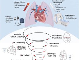
Abstract Purpose of review Acute pulmonary embolism (PE) is a heterogeneous disease process whose presentation varies widely between individuals who are asymptomatic, develop cardiogenic shock, or experience acute PE-related mortality...
Read More
Abstract In 2018, the position paper ‘Imaging the adult with congenital heart disease: a multimodality imaging approach’ was published. The paper highlights, in the first part, the different imaging modalities..
Read More

Mitral regurgitation (MR) is the second most common valve disease needing intervention in the Western world, after degenerative aortic stenosis.1 Echocardiography is the key examination in the assessment of MR patients.2 Echocardiography..
Read More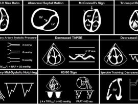
Introduction: Pulmonary embolism (PE) is a potentially fatal disease encountered in the hospital setting. Prompt diagnosis and management can improve outcomes and survival. Unfortunately, a PE may be difficult to diagnose..
Read More
Mohammed Zidan, MBBCH, M.Sc Cardiology (Cardiology, Echocardiography and interventional Cardilogy Specialist at Al-Azhar university), Cairo, Egypt.
Read More
Large cystic RA mass Mohammed Zidan, MBBCH, M.Sc Cardiology (Cardiology, Echocardiography and interventional Cardilogy Specialist at Al-Azhar university), Cairo, Egypt.
Read More
1. Modified 4chamver view due to all cardiac chamber dilation. 2. Huge LA due to sever mitral stenosis. 3. Sever rheumatic mitral stenosis with diastolic AML doaming. 4. Sever LA..
Read More
Severe Mitral Stenosis and Moderate Aortic Regurgitation Mohammed Zidan, MBBCH, M.Sc Cardiology (Cardiology, Echocardiography and interventional Cardilogy Specialist at Al-Azhar university), Cairo, Egypt.
Read More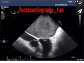
TEE shows Hugely dilated Left Atrium as a consequence of Rhematic Severe Mitral Stenosis, spontaneous Echo Contrast Grade IV “pre thrombus formation “ Mohammed Zidan, MBBCH, M.Sc Cardiology (Cardiology,..
Read More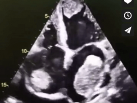
45 years old female patient with congestive Heart Failure “EF=22%” and history of stroke , the Echocardiography shows multiple big thrombi in the Heart chambers and moderate pericardial effusion ..
Read More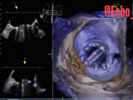
Severe Mitral Stenosis and Moderate Aortic Regurgitation Mohammed Zidan, MBBCH, M.Sc Cardiology (Cardiology, Echocardiography and interventional Cardilogy Specialist at Al-Azhar university), Cairo, Egypt.
Read More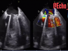
Paravalvular Leakage at 3 o’clock of Mitral Valve. Mohammed Zidan, MBBCH, M.Sc Cardiology (Cardiology, Echocardiography and interventional Cardilogy Specialist at Al-Azhar university), Cairo, Egypt.
Read More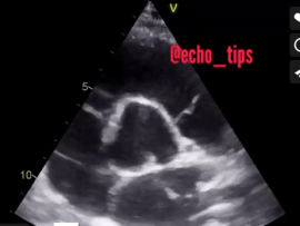
Mohammed Zidan, MBBCH, M.Sc Cardiology (Cardiology, Echocardiography and interventional Cardilogy Specialist at Al-Azhar university), Cairo, Egypt.
Read More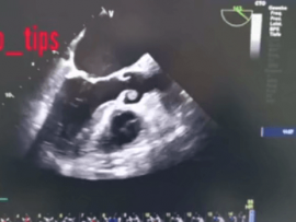
72 years ald female patient admitted to hospital because of delirium and hypertensive crises. In TTE we found clearly suspicious finding on the RCC of the Aortic Valve. For further..
Read More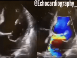
- Barlows prolapse. - Dilated left chambers, PMVL prolapse+ severe eccentric MR, Coanda effect, minimal pericardial effusion. - PML prolapse, severe MR eccentric jet, Dilated LA, Normal Lv systolic function.....
Read More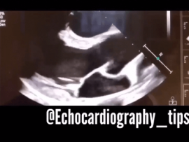
Mohammed Zidan, MBBCH, M.Sc Cardiology (Cardiology, Echocardiography and interventional Cardilogy Specialist at Al-Azhar university), Cairo, Egypt.
Read More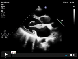
A 22-year-old man presented with palpitations and chest discomfort. Echocardiography revealed an enlarged, thickened mitral-valve leaflet with systolic prolapse of multiple segments (shown in a video), and a diagnosis of..
Read More
Parasternal short axis view, Apical four chamber view “A4C”. By placing the probe in the 5th to 7th intercostal space with a pointer directed to the lateral side till u..
Read More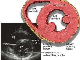
Parasternal short axis view. Papillary muscle level. The importance is ; 1) can assess the LV function 2)Ischemia “RWMAs” 3)VSD 4)Flattened D-shaped septum in Pulmonary embolism 5)LV wall thickness 6)Papillary..
Read More
Mohammed Zidan, MBBCH, M.Sc Cardiology (Cardiology, Echocardiography and interventional Cardilogy Specialist at Al-Azhar university), Cairo, Egypt.
Read More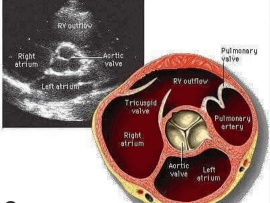
Beautiful illustrative picture about the parasternal short axis view, great vessel level Mohammed Zidan, MBBCH, M.Sc Cardiology (Cardiology, Echocardiography and interventional Cardilogy Specialist at Al-Azhar university), Cairo, Egypt...
Read More
Mitral Valve Leaflets and Scallops and its relation to Aortic Valve Sinuses Mohammed Zidan, MBBCH, M.Sc Cardiology (Cardiology, Echocardiography and interventional Cardilogy Specialist at Al-Azhar university), Cairo, Egypt...
Read More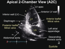
Mohammed Zidan, MBBCH, M.Sc Cardiology (Cardiology, Echocardiography and interventional Cardilogy Specialist at Al-Azhar university), Cairo, Egypt.
Read More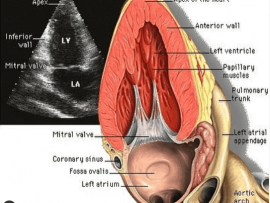
Apical 2 chamber view Importance: 1)Assessment of Ischemia “RWMAs” 2)Viewing LV apical thrombus 3)Assessment of MR and its mechanism 4)Assessment of pericardial effusion 5)viewing Coronary sinus and LAA ..
Read More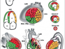
Different Echo views “A4C,A2C,PLAX and PSX” with the corresponding segment and its feeding vessel Mohammed Zidan, MBBCH, M.Sc Cardiology (Cardiology, Echocardiography and interventional Cardilogy Specialist at Al-Azhar..
Read More