Pierluigi Incarnato, TFCPC Cardiovascular and Abdominal Techinician Sonographer, Cardiac Physiologist at San Michele Nursing Home (Italy)
Read More
Aortic dissection is a serious condition in which there is a tear in the wall of the major artery carrying blood out of the heart (aorta). As the tear extends..
Read More
Apical thrombosis after previous acute myocardial infarction. Pierluigi Incarnato, TFCPC Cardiovascular and Abdominal Techinician Sonographer, Cardiac Physiologist at San Michele Nursing Home (Italy)
Read More
Nodule of Aranzio Anatomy of the aortic valve Pierluigi Incarnato, TFCPC Cardiovascular and Abdominal Techinician Sonographer, Cardiac Physiologist at San Michele Nursing Home (Italy)
Read More
Video Atrium Measurements Cardiovascular Sonographer Pierluigi Incarnato, TFCPC Cardiovascular and Abdominal Techinician Sonographer, Cardiac Physiologist at San Michele Nursing Home (Italy)
Read More
Pierluigi Incarnato, TFCPC Cardiovascular and Abdominal Techinician Sonographer, Cardiac Physiologist at San Michele Nursing Home (Italy)
Read More
[gallery ids="22031"] States that the volume of blood that passes through the mitral valve should be equal to the volume that passes through the aortic valve. SV = systolic range;..
Read More
In the last years, new trends on patient diagnosis for admission in cardiac intensive care unit (CICU) have been observed, shifting from acute myocardial infarction or acute heart failure to..
Read More
Apical akinesia in a patient undergoing primary PCI of residual IVA, with critical stenosis of CX and CTO of the right coronary. Pierluigi Incarnato, TFCPC Cardiovascular and Abdominal Techinician Sonographer,..
Read More
Severe mitral Stenosis insufficiency of the rheumatic type; doming of the flaps with hypomobility of the LMP. It can be seen Tunnel of the mitral inflow with minimal anatomical opening..
Read More
Background: McConnell's sign (right ventricular free wall [RV] hypokinesia with apical sparing on echocardiography) is often described as very specific for the diagnosis of pulmonary embolism (PE). In the video..
Read More
How to suspect pulmonary vascular disease ?! Here are some useful points: - Estimation of systolic pulmonary pressure - Pulmonary artery size and doppler - Right atrium / ventricle and..
Read More
Pierluigi Incarnato, TFCPC Cardiovascular and Abdominal Techinician Sonographer, Cardiac Physiologist at San Michele Nursing Home (Italy)
Read More
Case report Obstructive accessory mitral valve tissue in an adult patient. Background Accessory mitral valve tissue AMVT is a rare congenital Cardiac malformation that sometimes causes left ventricular outflow tract..
Read More
Pierluigi Incarnato, TFCPC Cardiovascular and Abdominal Techinician Sonographer, Cardiac Physiologist at San Michele Nursing Home (Italy)
Read More
Pierluigi Incarnato, TFCPC Cardiovascular and Abdominal Techinician Sonographer, Cardiac Physiologist at San Michele Nursing Home (Italy)
Read More
Giant aorta from chronic aortic dissection: The true light and false trombised light is appreciated. Furthermore it is possible to observe the breach of entrance which feeds and supplies the..
Read More
Type 1R Type 4LR Pierluigi Incarnato, TFCPC Cardiovascular and Abdominal Techinician Sonographer, Cardiac Physiologist at San Michele Nursing Home (Italy)
Read More
Pierluigi Incarnato, TFCPC Cardiovascular and Abdominal Techinician Sonographer, Cardiac Physiologist at San Michele Nursing Home (Italy)
Read More
Pierluigi Incarnato, TFCPC Cardiovascular and Abdominal Techinician Sonographer, Cardiac Physiologist at San Michele Nursing Home (Italy)
Read More
Pierluigi Incarnato, TFCPC Cardiovascular and Abdominal Techinician Sonographer, Cardiac Physiologist at San Michele Nursing Home (Italy)
Read More
Lipomatosis hypertrophy of the interatrial septum▫️ is a rare and benign cardiac pathology characterized by the accumulation of adipose tissue; according to some studies, performed on autopsy findings, it constitutes..
Read More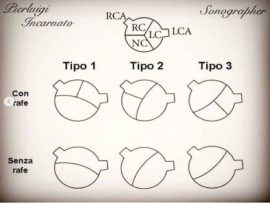
Bicuspid valve Type 2 without Rafe, with formation of bacterial endocarditis. Pierluigi Incarnato, TFCPC Cardiovascular and Abdominal Techinician Sonographer, Cardiac Physiologist at San Michele Nursing Home (Italy)
Read More
The takotsubo syndrome also called hypertrophic stress cardiomyopathy or shattered heart syndrome, is a clinical entity characterized by dysfunction of the left ventricle that manifests itself with symptoms that can..
Read More
Post-infarct patient with akinesia of the left ventricular apex, after about ten days from the first echocardium performed immediately after angioplasty, an important ecocontrast is evident in the left cavity..
Read More
Persistent left upper quarry in the long axis parasternal it is shown the dilated coronary sinus due to the persistence of the left superior vena cava, during the examination according..
Read More
Pierluigi Incarnato, TFCPC Cardiovascular and Abdominal Techinician Sonographer, Cardiac Physiologist at San Michele Nursing Home (Italy)
Read More
Not all masses visible at echocardiography are tumors intracardiac. The cardiac structures that can erroneously be interpreted as intracardiac masses are: • The Eustachian valve and the Chiari network •..
Read More
Pierluigi Incarnato, TFCPC Cardiovascular and Abdominal Techinician Sonographer, Cardiac Physiologist at San Michele Nursing Home (Italy)
Read More
Pierluigi Incarnato, TFCPC Cardiovascular and Abdominal Techinician Sonographer, Cardiac Physiologist at San Michele Nursing Home (Italy)
Read More