Type 1R Type 4LR Pierluigi Incarnato, TFCPC Cardiovascular and Abdominal Techinician Sonographer, Cardiac Physiologist at San Michele Nursing Home (Italy)
Read More
Pierluigi Incarnato, TFCPC Cardiovascular and Abdominal Techinician Sonographer, Cardiac Physiologist at San Michele Nursing Home (Italy)
Read More
Pierluigi Incarnato, TFCPC Cardiovascular and Abdominal Techinician Sonographer, Cardiac Physiologist at San Michele Nursing Home (Italy)
Read More
Pierluigi Incarnato, TFCPC Cardiovascular and Abdominal Techinician Sonographer, Cardiac Physiologist at San Michele Nursing Home (Italy)
Read More
Lipomatosis hypertrophy of the interatrial septum▫️ is a rare and benign cardiac pathology characterized by the accumulation of adipose tissue; according to some studies, performed on autopsy findings, it constitutes..
Read More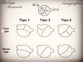
Bicuspid valve Type 2 without Rafe, with formation of bacterial endocarditis. Pierluigi Incarnato, TFCPC Cardiovascular and Abdominal Techinician Sonographer, Cardiac Physiologist at San Michele Nursing Home (Italy)
Read More
How do we know something is normal if we haven't seen the abnormality first? Normal MV vs a metallic MV replacement. The echo lady; Lorena De Vanna, is a..
Read More
Are most commonly seen between six and 10 days following an acute myocardial infarction (MI). They occur at the left ventricular apex and are more common following an anterior wall..
Read More
Aortic dissection is when the weakened wall of the aorta tears, causing blood to leak between the layers that makes up the wall. This can happen suddenly or slowly over..
Read More
A dilated aortic root is when the first section of the aorta, where the aortic valve resides, becomes enlarged. When this enlargement reaches a critical size, there is a risk..
Read More
Normal aortic valve opening properly Vs a heavily calcified aortic valve with severe stenosis. The echo lady; Lorena De Vanna, is a cardiac and respiratory physiologist graduated from the..
Read More
Left ventricular (LV) hypertrabeculation is defined by the presence of three or more trabeculations apically and up to the level of papillary muscles, seen in one echocardiographic view. It can..
Read More
The echo lady; Lorena De Vanna, is a cardiac and respiratory physiologist graduated from the Central University of Venezuela. She currently holds British Society of Echocardiography accreditation and works..
Read More
52 years old male patient with constrictive pericarditis after a CABG in 2018. Constrictive pericarditis is a potentially curable condition caused by a variety of situations which result in inflamed,..
Read More
Septal Occluder is a percutaneous, transcatheter, atrial septal defect closure device intended for the occlusion of atrial septal defects (ASD). ⠀ Patients indicated for ASD closure have echocardiographic evidence of..
Read More
Abstract Accurate echocardiographic evaluation of the systemic is challenging because of its specific morphology and contraction patterns. We present a detailed multimodality assessment of the systemic right ventricle, analyze the relative..
Read More
The echo lady; Lorena De Vanna, is a cardiac and respiratory physiologist graduated from the Central University of Venezuela. She currently holds British Society..
Read More
The echo lady; Lorena De Vanna, is a cardiac and respiratory physiologist graduated from the Central University of Venezuela. She currently holds British Society..
Read More
En este video les muestro como medir la aorta en diferentes niveles. In this video i will show you how to measure the Aorta at different levels. The..
Read More
El TAPSE es una medida que utilizamos para evaluar la funcion sistolica del ventriculo derecho. This is a parameter we use to assess the right ventricular systolic function. ..
Read More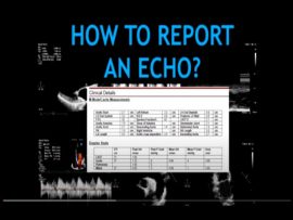
En este video les muestro como crear tu reporte de ecocardiograma. Esto es de acuerdo a los requerimientos minimos que debe tener un reporte. In this video I am..
Read More
Measuring the Right atrium Right Ventricular strain Left Ventricular strain The echo lady; Lorena De Vanna, is a cardiac and respiratory physiologist graduated from the Central University of..
Read More
Como medir el diametro de la auricula izquierda. Recuerda indexar todas tus mediciones por el area de superficie corporal. How to properly measure the Left Atrium diameter. Always remember..
Read More
Como medir el volumen de la auricula izquierda por el metodo de Simpsons. How to measure the left atrium volume by Simpson Biplane method. The echo..
Read More
Como medir el tracto de salida del ventriculo izquierdo (TSVI) acorde con la Sociedad Britanica de Ecocardiografia (BSE). Si tienes algunas dudas, por favor siempre chequea los guidelines en tu..
Read More
Video basico para explicarles la estructura de un ecocardiograma y en proximos videos voy a demostrarles como obtener las diferentes vistas (Yay). This is a basic video so you..
Read More
The echo lady; Lorena De Vanna, is a cardiac and respiratory physiologist graduated from the Central University of Venezuela. She currently holds British Society of..
Read More
Parasternal short axis view at the Aortic valve level: - RVTO: right ventricular outflow tract. - PV: pulmonary valve. - PA: pulmonary artery. - AV: aortic valve...
Read More
. The echo lady; Lorena De Vanna, is a cardiac and respiratory physiologist graduated from the Central University of Venezuela. She currently holds British Society..
Read More
The echo lady; Lorena De Vanna, is a cardiac and respiratory physiologist graduated from the Central University of Venezuela. She currently holds British Society of..
Read More