Pulsatil and NonPulsatil Current in Cardiopulmonary Bypass
Abstract
Heart disease is among the most important health problems in our country and in the world. Heart diseases are widely repaired in adults, children and newborns with open heart surgery techniques. Within the scope of cardiac surgery; heart valve repairs, coronary artery diseases, heart and lung transplants can be performed.
Cardiovascular surgery requires a complicated surgical technique compared to other surgical branches. In order to perform open heart surgery, the heart must be ane bloodless and immobile. Therefore, a technique called extracorporeal circulation has been developed.
In open heart surgery, the pump functions of the heart and the gas exchange functions of the lungs are performed outside the body and with the help of different mechanical equipment is called extracorporeal circulation. Extracorporeal circulation technique is often indispensable in terms of surgical comfort and safety.
Open heart surgery has made significant progress since John Gibbon’s first successful cardiopulmonary bypass in 1953.
Extracorporeal circulation has been an important turning point in the development of open heart surgery. In this way, open heart surgery was successfully performed on many patients around the world. This system ensures the maintenance of the operation in a bloodless area without disrupting the perfusion of the vital organs of the body during open heart surgery.
Due to the use of extra-body circulation, research is underway to reduce morbidity and mortality rates for this operation, which is basically non-physiological, and a cardiopulmonary bypass machine constitutes an important part of the studies.
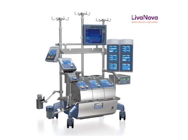
Extracorporeal circulation (EKD) is a system based on the principle that the patient’s blood is taken from the main veins of the heart or directly from the right atrium and oxygenated and given back to the patient through the patient’s main arteries. In order for this system to occur, there must be some equipment. These are mainly heart lung machine, oxygenator reservoir, tubing set cannulas, oxygen mixer, heater cooler priming solutions, cardioplegi solutions.
However, this circulatory system differs from physiological circulation. The heart provides physiologically pulsatil current. Extracorporeal circulation provides nonpulsatil current. With the developing technology in recent years, machines that provide pulsatil current have been developed in many clinics. Especially after aortic clamping was placed, cardiac arrest occurred and pulsatil current circulation started to be provided.
The cardiopulmonary bypass machine provides pulsatil or non-pulsatil current with the main pump. In principle, the physiological pulsatil current is thought to be superior to non-pulsatil current. However, the debate about the superiority of these two movements towards each other has been going on for a long time.
Perfusion support provided by heart lung machine and components is of vital importance for the patient. During cardiopulmonary bypass, perfusionists should evaluate many parameters and intervene immediately and should not ignore any issues that may pose problems.
Since the birth of the idea of extracorporeal circulation, many researchers have focused their studies on the current dynamics during perfusion and examined its effects on the organism.
Some of the researchers argued that in pulsatil current applications, organ blood supply was better than flat current; others reported that they did not detect significant functional differences between the two currents.
There is no consensus yet on the effects of this two-way current shape, which has different physical characteristics, on organ functions.
Pumps
Various pump types are available for cardiopulmonary bypass (KPB). The ideal pump for KPB should have the following characteristics:
-
It should be able to pump blood at a current speed of 7 lt/min against pressure of 500 mmHg.
-
Pumping movement should not damage the flooded and aceullar elements of the blood.
-
All surfaces that come into contact with blood flow should be continuous, there should be no dead cavities that can cause stagnation or turbulence, they should be disposable and should not contaminate the permanent parts of the pump.
-
Pump current calibration must be complete for accurate moniterizing of blood flow.
-
In case of power failure, the pump must be operated manually.
-
It should be simple and safe to use.
Roller Pumps
It is the most commonly used pump in KPB in the last fifty years. They are easy-to-use, safe and relatively low cost pumps. Its popularity has decreased recently as a result of advances in systems using sentrifugal pumps.
The roller pump was first patented by Porter and Bradley in 1855. In 1877, Allen designed a pump for blood transfusion. In 1934, DeBakey et al. developed the Porter Bradley infusion pump with a modification. In 1959, Melrose further developed the design.
The cylindrical structure in the two rotating arms compresses a thick tube carrying blood through the pump tank, pushing the blood and thus producing continuous blood flow. The pump current depends on the number of turns per minute (rpm), the diameter of the tube and the length of the path where the pressure is applied.
Pumps rollers according to the number of cylinders; single, double and numerous. It was used in KPB in the 1950s and early 1960s with a single-cylinder pump. It consisted of a circular chamber and the tube was placed to loop 360°. Double cylinder pumps are the most widely used pumps for KPB. It consists of a semicircular rotary surface of 210° and cylinder arms with 180° separate placements. When one cylinder finishes its own occlusive phase, the occlusive phase of the other begins. Since one of the two cylinders presses continuously on the tube, the double roller pump produces relatively non-pulsatil current. Although multi-cylinder pump models are recommended, they do not have validity in clinical practice as they cause excess hemolization.
An important concept in roller pumps is the amount of occlusion. Occlusion, which describes the degree of pressure, can be adjusted by increasing or reducing the pressure of the cylinders to the tube. Occlusion is important because excessive compression causes hemolysis and wear of lines, while on the contrary, low occlusion can aggregate hemolysis, and more importantly, reduce forward current. The current provided by the roller pump may vary during KPB, and the change in occlusion throughout the bypass creates difficulty in determining the actual current.
The occlusion adjustment can be done as follows: by keeping the outflow line upright, the occlusion is gradually reduced so that the liquid level is about 60-75 cm above the pump and the liquid level drops by 1 to 12 cm per minute.
Centrifugal Pumps
In 1960, Saxton and Andrews designed the centrifugal pump for the first time. Sentrifugal pumps for KPB have been available since 1976. In many institutes, sentrifugal pumps have been routinely used in heart surgery instead of roller pumps for KPB. They are also used for mechanical circumvention support, ventricular assist, percutaneous cardiopulmonary support and extracorporeal membrane oxygenation.
Sentrifugal pumps, also known as rotadynamic or radial current pumps, move to the blood thanks to their fast rotating structure (blade, impeller, cone). Sentrifugal pumps are kinetic pumps that activate blood with the principle of artificial vortex created by an electric motor. The centrifugal force consisting of a cone rotating in the electromagnetic field rotates the blood from the patient through the inlet tube and moves the blood out of the chamber and is sent to the patient in the form of a nonpulsatil current from the outlet tube in this section.
Rotational movement of blood creates high current with low pressure. Afterload provides dependent current, and afterload changes are effective in the performance of the system. In the face of increased resistance due to sudden bends in the lines, the current decreases and separations or explosions in the pump lines are prevented. When the pump stops or slows down, the current rotates back (retrograde) from the arterial line to the pump. This creates a hemodynamic siphon effect and can cause air to enter the system from the cannulation zone. Therefore, when the pump stops, the arterial line should be clamped. In order not to encounter this problem, one-way valve systems have been developed for the arterial line and it is recommended to use it. In fact, when the air enters, the pump stops when the constive forces between the blood layers disappear. In addition, it is an important advantage of the pump to reduce the risk of embolism by collecting micro-air embolisms in the center of the vortex.


Advantages and Disadvantages of Rollers and Sentrifugal Pump

NIRS

NIRS was found in 1970; In 1985, it was first used by Ferrari and his friends to perform cerebral measurements in humans. With the approval of the American food and drug agency, it was produced in various brands and models and started to be used in the clinic in 1993.
A number of parameters used during standard monitoring (such as arterial blood pressure, arterial oxygen saturation) may not always be sufficient. Therefore, as a result of the studies, nirs method that shows tissue oxygenation more effectively, measures cerebral reginal saturation and is used noninvasively has been found. While other methods measure arterial blood pressure, NIRS reflects the longer-term effects of the changes. It is an increasingly accepted method.
This method benefits from the principle that light in the near infrared spectrum penetrates into the tissue, including bone and muscle. A prop is placed in the desired area for NIRS to measure. This propane has a light source, two light sensors. One of the sensors measures skin tissue, that is, more superficial, and the other measures bone and brain tissue, i.e. deep tissue oxygen.
Sensors are placed at constant distances from a light emitter, and algorithms extract superficial light absorption from deep absorption to provide an index of tissue oxygenation. The infrared beam transmitted by nirs device in cerebral recitation oxygen saturation measurement is said to show the saturation of the area called water sheed zone, which consists of a 75-85% venous and 15-25% arterial mixture at a depth of 1-1.5 cm, and is also used as somatic (liver, kidney, mesentery) in NIRS pediatric patients.

Impact of Pulsatil Current Selection on Cerebral NIRS
Various methods have been developed and successfully carried out over the years to minimize the harms of cardiopulmonary Bypas. The brain is one of the most susceptible organs to ischemia during cardiopulmonary bypass. The brain responds to changes in average arterial pressure by keeping cerebral blood flow constant. As long as the average arterial pressure is between 60-150 mmHg, the blood flow in the brain may remain constant. One of the most important causes of mortality after cardiac surgery is neurological complications. Neurological complications may occur at values below or above this pressure range.
Pulsatil current in roller pumps was first used especially in the 1980s. In Wesolowski’s study, a current speed of 130-200 ml/kg/min was maintained and pulsatil circulation had no additional advantage. Pulsatil current is one of the negative factors in which the roles hemolimate blood cells above normal in the pumps; Another reason for the under-preference is the high pressure that artificially generated pulsatility creates above a certain rate of the standard in the aortic cannula along the arterial line with oxygenator input and output.
Studies by Hornick and Taylor at KPB have predicted that pulsatil perfusion may be more beneficial in some cases; Pulsatil study was suggested to be more effective in patients with high risk of myocardial ischemia and infarction, patients with carotid artery symosis, patients with chronic renal failure and liver failure, and patients with arterial hypertension.
Renal damage at certain rates after cardiopulmonary bypass is a common complication. Many pharmacological and non-pharmacological kidney protective methods have been tried to minimize the negative effects of KPB on the kidney. One of these methods is the application of circulation in the form of pulsatil current, in accordance with the nature of the body during KPB.
According to the biochemical data of the study carried out by Ozturk S., no statistically significant difference was detected in two pulsatil and nonpulsatil patient groups. LDH, AST, ALT, direct and indirect bilirubin levels tended to increase in patients with two types of currents. However, liver function tests have detected an increase; rather than the current type, it has concluded that it develops as a complication due to KPB. They concluded that these effects are temporary due to the liver’s ability to repair and regenerate itself.
Blood gas lactate and pH measurements are among the routine applications to evaluate adequate oxygen delivery. Louagie et al.’s study observed some increase in blood lactate levels during and after KPB, but was not affected by the pulsatil or nonpulsatility of this current.
Cerebral autoregulation cannot be achieved if the average artery pressure drops below 55-60 mmHg at normal temperature. In hypothermia, the brain uses oxygen more efficiently because the metabolic rate decreases. Excitator substances released from ischemic neurons also increase hypothermia. In ozturk S.’s study, there was no statistically significant difference in cerebral NIRS measurements in pulsatil and non-pulsatil groups in adult patients who had coronary artery bypass surgery. Another study with the NIRS device alone has shown that pulsatility does not alter cerebral oxygenation.
Neurological problems are among the biggest causes of postoperative mortality and morbidity in patients undergoing KPB procedure. In samet D.’s study, there was no statistically significant difference when both groups were encountered in terms of their release from intensive care and discharge from the hospital. However, statistically significant differences were found in terms of intensive care ventilation time by group (p=0.040). It was observed that the intensive care ventilation time of patients who applied non-pulsatil current was statistically significantly higher than that of the pulsatil current patient group.
As a result, the superiority of non-pulsatil current and pulsatil current over cerebral NIRS could not be observed. However, it was observed that lactate levels were significantly lower in patients with pulsatil current according to postoperative 24th hour arterial and venous blood gas results, as well as; Patients who applied pulsatil current in terms of ventilation time were found to be extubated earlier. However, it was observed that this did not significantly affect the intensive care stay and hospital stays of both groups of patients.
Despite the natural protective autoregulation mechanism, the brain is susceptible to injury during KPB. During KPB, some factors affecting the brain, including temperature, blood pressure, blood vizcosity, oxygen and carbon dioxide pressure, have been shown. In addition to maintaining the function of cerebral activity, pulsatil current has the effect of preventing cerebral asidose encountered in the early stage of bypass with non-pulsatil current. Kono et al. showed a 25% difference in cerebral vascular resistance measured in non-pulsatil and pulsatil patient groups. This important difference in cerebralvascular resistance may be responsible for improving regional blood flow and distribution, reducing excess cerebral lactate. Anaerobic metabolism can be prevented by applying pulsatil blood flow and the continuity of regional blood flow can be ensured. Other studies focusing on cerebral oxygen consumption, especially during the critical cooling and heating stages of the operative process, show that pulsatil flow is associated with increased cerebral metabolism in pulsatil flow conditions. Some researchers suggest that there is no difference between clinically applied pulsatil current and non-pulsatil flow in terms of cerebral effects.
Hemodynamic Effects of Pulsatil Current
The hemodynamic effects of pulsatil KPB are the fastest and most pronounced for the clinician. One of the most reported clinical findings in this regard is the effect of the flow type on SVR, that more physiological condition is maintained in patients with pulsatil flow regimen. Taylor (1980) confirmed this effect when he showed that patients under non-pulsatil KPB developed progressive vasoconstruction. It found that if left unchecked, cardiac surgery could seriously endanger the patient in the post-perfusion stage.
Vasoconstruction, which develops with non-pulsatil current, often needs to be reversed by pharmacological intervention in the critical end-of-surgery phase. However, Taylor (1980) showed that the use of pulsatil current during KPB was an alternative strategy to pharmacological intervention in the approach to unexpected postoperative complications. Despite verifiable inferences that vasoconstruction effects are successfully mitigated by pharmacological intervention, it is clear that preventing this using pulsatil current is a more satisfactory and physiological solution than pharmacological treatment.
Numerous factors have been identified, including system activation of renin-angiotensin and catecholamine release vasopressin and local tissue vasoconstructors, which contribute to vasoconstruction associated with non-pulsatil current. However, in order to understand why these different current methods produce different responses in patients with KPB, it is necessary to go over the fundamental differences between the two models in terms of sending blood to the tissues, and it should be borne in mind that the opinion is divided regardless of this simple mechanism.
Effects of Pulsatile Blood Flow on Microcirculation
Burton (1954) suggested that while arterial blood pressure weakens after systole, the blood flow in the microcirculation continues until a certain critical closing pressure is reached in the precapillary arterioles, and when the pressure builds up, the blood flow in the capillaries will stop at this point. It also showed that pulsatility increases capillary patency period and blood flow.
This view may largely explain many of the perceived benefits of pulsatile blood flow as the preferred type of flow. As early as 1938 McMaster and Parsons showed that lymph flow is greatly reduced when blood flow is pulseless.
Ogata et al. (1960) confirmed that capillary blood flow and diameter are affected by the presence of a pulse, regardless of total blood flow and mean pressure. At the same time (Takeda 1960) showed that there is widespread collapse of the capillary structure in the presence of non-pulsatile flow in the capillary structure, with the association of decreased blood flow and increased capillary shunt. In these repeated animal experiments, mean blood flow and arterial pressure are the same in both pulsatile and non-pulsatile flow models, suggesting that the presence of a pulse itself is responsible for these demonstrated benefits. Capillary diameter reduction and capillary transit are associated with clinical signs of increased peripheral vascular resistance in non-pulsatile flow. It has recently been suggested that pulse pressure may not be responsible for the maintenance of peripheral patency in the presence of pulsatile blood flow. Shepard et al.
Using both mathematical and physical modeling, they determined that the pulsatile blood flow model contains up to 2.4 times the increased energy of the non-pulsatile flow model at the same mean arterial pressure and flow. Wilcox et al. (1970) confirmed that the pulsatile current regime is responsible for the distribution of significantly greater energy to tissues as determined. Previously, Shepard et al. (1966) suggested that more energy provided in the presence of pulsatile blood flow is responsible for the maintenance of capillary opening and is responsible for fluid exchange at the extracellular level, which can be seen at the capillary level, thus ensuring the continuity of cell metabolism.
Using a rabbit model, Parsons and McMaster (1938) noted that the isolated ear preparation developed edema when perfused with non-pulsatile blood flow, and those perfused with the pulsatile flow regimen remained essentially normal. These findings were confirmed by Shepard et al. tended to add weight to their findings. Prior et al. (1955) suggested that the pulse is responsible for fluid exchange at the capillary level and the mean arterial pressure is responsible for maintaining fluid balance.
The structure of the pulse pressure profile at the capillary level, the mean blood pressure, the osmotic pressure in the blood and between the cells, the nutrient exchange at the cell level and the continuation of the fluid balance, which is mentioned in this simple relationship previously described by Prior as «pulsed reverse osmosis» (pulsed reverse osmosis). are responsible factors. This is largely in line with the Starling Principle (1896), except that Starling did not realize the importance of the presence of a pulse and the different pore distribution at the capillary level. (Prior et al.1996) The effect of pulsatile current on this microcirculation may be the basis for its importance in maintaining normal physiological functions. The support provided by this theory draws attention to ongoing laboratory and clinical studies.
In a prospective study, 20 high-risk cardiac surgery patients were followed during CPB with 10 pulsatile streams and 10 non-pulsatile streams, changes in sublingual mucosal microcirculation were evaluated with orthogonal polarization spectral imaging together with near-infrared spectroscopic indices of thenar muscle oxygen saturation and scoring. It was concluded that a change in microvascular blood flow occurred over time, with the pulsatile group maintaining normal perfusion characteristics while the non-pulsatile group showed impaired perfusion during CPB.
Metabolic Effects of Pulsatile Perfusion
The presence of pulse has been the focus of research and an important subject of academic literature for decades because of its importance for metabolic processes. Most of the major organ systems have been studied in this respect, and the effects of pulsatile current on their functions and structures have been extensively studied.
Pulsatil Current and Kidney
In 1889, Hamel (1889), using isolated kidney preparations, confirmed that the pulse had a significant effect on kidney function. This was supported by Gesell’s (1913) study, which found that improved kidney function associated with pulsatil blood flow was the result of better gas exchange at the capillaries level, which was provided by increased lymph current. It has been shown that the pulse itself is associated with maintaining kidney function, in isolated kidney preparation, revealed by Kohlstaedt and Page (1940), the absence of pulse in blood flow is linked to an increase in renin release and a decrease in urine output. These early findings formed the basis of an important working group in the coming years. These results were later made by Mavroudis (1978) in his excellent re-review of the subject, which stated that there were those who objected to the importance of pulsatil current. Selkurt (1951), Ritter (1952) and Goodyer and Glenn (1951) all found that kidney function was not affected by the presence of a pulse in isolated kidney preparations, provided that the average blood pressure was maintained at the physiological level. Differences between the modeling methods used in these studies may be partly responsible for conflicting results. However, overwhelming studies using various KBP models tend to support early findings that show that pulsatil current and renal functions are maintained to some extent in isolated organs.
Using animal KPB models, fintersbuch et al. (1961) and Nakayama et al. (1963) maintained renal venous rotation in the presence of pulsatil blood flow and some loss in normal renal artery configuration was associated with non-pulsatil current. Using a similar model, Barger and Herd (1966) linked these findings to changes in intraceldone blood flow and reduced sodium excretion. Apart from this, the effect of pulsatil blood flow on kidney blood flow was also used by Boucher et al .1974 in pulsatil blood flow conditions, micro spheres marked radioactively in the bypass model to show that kidney blood flow was maintained. This effect was also described by Mori et al. (1988). In fact, when dogs were exposed to pulsatil blood flow during the reperfusion period after the hypothermic circatatuar arrest period, the renal blood flow was quite high and a rapid and complete improvement in kidney function was found. Nakamura and ark (1989) used pulsatil and non-pulsatil KBP in the same dog models, renal blood flow, lactate extraction, urine output were higher in favor of pulsatil current. Undar et al. (1999) used a piglet model to show once again that organ blood flows, including blood flow to the kidney, are preserved in pulsatil current conditions (30). Most of the findings from isolated organ and animal studies have been confirmed in clinical trials. German et al. (1972) showed that non-pulsatil blood flow was associated with the onset of renal hypoxia and acidosis faster than pulsatil current, despite seemingly sufficient total blood flow. Mukherjee et al (1973) confirmed these findings. They reported a decrease in po2 in the medulla in the presence of non-pulsatil current, increased levels of local lactate level and decreased oxygen intake. Taylor et al ( 1982) announced results associated with the use of pulsatil blood flow in open heart surgery, decreased plasma angiotensin levels and increased urine production (33). Similarly, Landymore et al. (1979) found that urine output increased with pulsatil current and plasma renin levels were higher with non-pulsatil current. Findings in adult cardiac surgery have also been useful in pediatric surgery. Williams et al ( 1979) infants found that the amount of urine in patients exposed to pulsatil blood flow was twice as high as in non-pulsatil flow regimen.
In line with animal and laboratory studies, not all clinical findings resulted in favor of pulsatil current. A number of studies indicate that there is no real difference between pulsatil and non-pulsatil flow regimes in terms of renal function. Badner et al ( 1992) found that the flow regimen had little or no effect on renal function, concluding that pulsatil blood flow in routine KPB patients was related to reduced urine output and creatine clinching. These results may be associated with a number of variables, including the pulse structure and anesthesia regimen, which have been shown to affect clinical findings. Anesthesia regimens with different hemodynamic effects themselves, different pump output profiles may provide an explanation for the discrepancy in the findings regarding the physiological effects of pulsatil current during KPB. Until the ideal blood flow profile and anesthesia regimen reach some degree of consensus, discussions around pulsatil flow can continue.
In a paper published in the Turkish Journal of Thoracic Cardiovascular Surgery (TKDC) in 2013, 40 patients over the age of 70 were divided into two groups and compared to the pulsatil and non-pulsatil current regimen. In this study, pre-operative, post-KPB and post-op 3. Day creatinine, statin C, urea nitrogen values and urine output during KPB were looked at. In the pulsatil current group, statin C, creatinine, blood urea nitrogen values were found to be relatively low in the 3rd day following surgery, and it was suggested that pulsatil current was a simple and safe method in KPB and could prevent acute renal failure (ABY) in the elderly.
In another study, 10 patients were divided into two groups: pulsatil and 12 nonpulsatil during KPB. Cortisol levels were studied to investigate the effect of the current pattern on the adrenal cortex, and after the correction to eliminate the effect of hemodilution, it was determined that cortisol levels were significantly higher in patients undergoing pulsatil perfusion compared to the other group, and in the light of these findings, it was suggested that pulsatil blood flow was more physiological in terms of adrenal cortex functions.
In a meta-analysis, 477 patients were given non-pulsatil perfusion during KPB, and there was not enough evidence to show a difference in average postoperative creatinine and urea values between the groups, but the pulsatil perfusion group had significantly higher creatinine clereyne and lower serum lactate levels. This study found great variability between pulsatil perfusion studies, and effective pulsatiity creation and evaluation methods on bypass differed greatly between the articles. This analysis shows that pulsatil perfusion during CPC is useful in kidney protection and should be taken into account.
Pulsatil Blood Flow and Its Effects on The Liver and Pancreas
The interest in the effect of KPB on pancreatic function was initiated by early, sporadic and isolated findings with increased plasma amylase levels associated with non-pulsatil bypass, and Feiner (1976) reported 16% of patients who had open heart surgery under non-pulsatil current conditions. Baca et al. (1979) found that in the dog model, pancreatic function was maintained under pulsatil blood flow conditions and deteriorated under non-pulsatil conditions. Saggau et al. (1980) monitored insulin levels with glucose, glucogan and growth hormone, concluding in human and animal studies that during the perfusion phase of the operation, pulsatil blood flow maintained normal pancreatic functions. Mori et al. (1988) concluded that pancreatic function was maintained in 24 dogs perfused under both hypothermia and normothermia, only in the presence of pulsatil current.
Examination of liver function during pulsatil and non-pulsatil KPB gave similar results to pancreatic functions. Pappas and ark (1975), who used serum glutamic-oxayloacetictransaminase (sGOT) as an indicator of liver damage, found that pulsatil blood flow was associated with maintaining hepatic function. Mathie et al. (1984) demonstrated that pulsatil KPB in dogs maintains both hepatic blood flow and function. Mathie et al. (1984) reported that liver blood flow reacted typically vasoconstructively during non-pulsatil current KPB and showed a decrease in hepatic oxygen consumption. Further protection of physiological blood flow in the liver and pancreas with pulsatil current is the most important factor in maintaining normal function and structure in these tissues.
Pulsatil Blood Flow and Intestines
Abdominal complications caused by KPB constitute an important part of reported operative mortality. In a study of 500 open cardiac surgery procedures, Gauss et al. (1994) reported that 1.8% of patients exhibited some abdominal cramping. Baue (1993) confirmed in this extremely important review of 5924 patients that gastrointestinal problems cause a significant amount of KPB-related mortality and morbidity, and suggested that the mechanism that led to these complications was mesenteric hypoperfusion, which occurs in the presence of non-pulsatil current. Many studies have linked intestinal ischemia and endotoxemia due to this hypoperfusion. (Bowles et al 1995) Endotoxemia associated with KPB in children has been the focus of research for some time. Anderson and Baek (1992) associate high levels of endotoxins in children with increased intestinal permeability due to mesenteric ischemia. Anderson et al. (1987) has previously shown that endotoxemia during and after bypass is not associated with preoperative infection. In a series of studies, peak levels of endotoxin are shown in the heating phase after opening the aortic cross-country clamp. Ohni et al. (1994) confirmed the importance of the warming phase in the pathophysiology of all 25 endotoxemias using dogs in the bypass model. These authors showed that there is a difference between mesenteric oxygen presentation and consumption at this stage of the procedure and that there is an increase in intestinal permeability associated with it. Tao et al (1995) used a pig model to show that the intestinal mucosa actually became ischemic during KPB, suggesting that this was due to two factors in reperfusion in the form of altering blood flow and tissue oxygen demand. Riddington et al. (1996) confirmed these findings in the clinical model. It revealed that there was an increase in ischemia and permeability in the bowel mucosa in KPB patients and that endotoxin was detected in plasma in 42% of these patients. These studyers also found that increased intestinal pH did not return to normal until the non-pulsatil flow regimen ended and the heart circulation took over. Later, Hamulu et al. (1998) described a similar effect.
Fiddian-Green (1990) suggests that pulsatil blood flow can lead to increased blood flow in the intestine, decreased mucosaous ischemia and increased oxygen intake. He also recommended preoperative intestinal cleansing and parenteral antibiotics as a prophylactic approach to reduce the incidant of endotoxemia. Reilly and Bulkey (1993) argued that the vasoactive response to circulatory shock in the intestine was mediated by the activation of the renin-angiotensin system, which leads to intestinal hypoperfusion and ischemia, which causes an increase in intestinal permeability. Further physiological circulation structure and pulsatil current use in maintaining current (Taylor et al.1979b) can be associated with low renin-angiotensin activation level, which can be associated with a decrease in the incidance of intestinal complications.
Factors limiting the effectiveness of pulsatil blood flow
Mechanical pulsatil pump systems are efficient machines that can provide blood flow with significant hydraulic power. However, the effectiveness of these devices is limited to a number of external factors in clinical practice. In routine clinical practice, the arterial pump is positioned far away from the aorta, ideally the pump should be placed near the aorta, as with the native heart, but unfortunately in traditional clinical perfusion circuits this is unrealistic. The pump and the aorta often include devices such as membrane oxygenators and arterial filters with large surface area, and a highly flexible pipeline. The movement and yawning of the tubing and the absorption of all energy by the membrane surfaces reduces the effectiveness of the pulse manufacturer. Many studies have identified energy absorption properties in frequently used clinical circuits. However, the most restrictive factor in terms of pulsatil blood flow is aorticcanul. The diameter of the aorticcanul is usually less than 4 or 5 mm, after the arterial line of 8-10 mm there is a significant descent. This diameter reduction has a pronounced effect on the pulse structure sent to the aorta. Some researchers have shown that it is possible to produce pulsatil flow in physiological proportions in the aorta through a large cannula. (Runge et al.1992) But the placement of large-scale cannulas in the aorta is an unacceptable risk for an already complex surgical procedure.
In a study on the benefits of pulsatil current, important factors related to why this debate has continued over the last 55 years have been tried to be clarified, it is emphasized that not only the pump is important to produce sufficient quality pulsatility, but also the geometry and internal diameter of the oxygenator, arterial cannula. Accordingly, the pump should be examined in terms of hemodynamic energy production, the advantage of pressure drop in the selection of oxygenators should be taken into account, there should be no hemodynamic energy loss when passing through the membrane. The membrane called «Flatsheet» significantly reduces pulsatility. In the new generation of oxygenators with integrated arterial filters, the advantages of pressure drop as well as micro embolism retention capacities are the preferred reason. In this study, it was observed that the internal diameters studied with eight different arterial cannulas with a diameter of 10 fr differed from 2.08 mm to 2.69 mm, and the line pressure changed dramatically at the same current. This not only reduced the pulsatil energy sent, but also caused an artificial increase in line pressure. They proposed the formula «Energy equvalent pressure» (EPP) to measure the amount of pulsatil current. In the light of this data, comparisons between perfusion modes should be made after these basic steps are taken.
The Future of Pulsatil Perfusion
The development of pulsatil perfusion was in line with its technology, which developed in the last century. Pulsatil roles have a radical impact on the acceptance and clinical use of pulsatil flow systems that are part of the pump. Advances in pump design have led to the production of pulsatil pumps that can produce pulse pressure or current profile close to physiology, as shown in laboratory studies. However, the limiting factors described in the previous section still persist today. Recent developments in clinical KPB offer some hope for the future of pulsatil perfusion. The main limiting factor for the reduction of the perfusion circuit and the requirements for the perfect optimization of pulsatil KPB is the arterial cannula, making the entire perfusion device closer to the patient. It remains a serious obstacle to pulse transmission. If this cannulation problem can be addressed, it may be possible to achieve all the perceived benefits of real physiological pulsatil flow in the near future.
Author Note
This article was prepared by compiling from the articles in the bibliography. It is not intended for any benefit or gain. Our main goal is the dissemination of knowledge. If you are requesting that your article be removed from the article, you can contact heparinsodyum@yahoo.com.