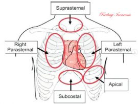
How to suspect pulmonary vascular disease ?! Here are some useful points: - Estimation of systolic pulmonary pressure - Pulmonary artery size and doppler - Right atrium / ventricle and..
Read More
Pierluigi Incarnato, TFCPC Cardiovascular and Abdominal Techinician Sonographer, Cardiac Physiologist at San Michele Nursing Home (Italy)
Read More
Case report Obstructive accessory mitral valve tissue in an adult patient. Background Accessory mitral valve tissue AMVT is a rare congenital Cardiac malformation that sometimes causes left ventricular outflow tract..
Read More
Pierluigi Incarnato, TFCPC Cardiovascular and Abdominal Techinician Sonographer, Cardiac Physiologist at San Michele Nursing Home (Italy)
Read More
Pierluigi Incarnato, TFCPC Cardiovascular and Abdominal Techinician Sonographer, Cardiac Physiologist at San Michele Nursing Home (Italy)
Read More
Giant aorta from chronic aortic dissection: The true light and false trombised light is appreciated. Furthermore it is possible to observe the breach of entrance which feeds and supplies the..
Read More
Type 1R Type 4LR Pierluigi Incarnato, TFCPC Cardiovascular and Abdominal Techinician Sonographer, Cardiac Physiologist at San Michele Nursing Home (Italy)
Read More
Pierluigi Incarnato, TFCPC Cardiovascular and Abdominal Techinician Sonographer, Cardiac Physiologist at San Michele Nursing Home (Italy)
Read More
Pierluigi Incarnato, TFCPC Cardiovascular and Abdominal Techinician Sonographer, Cardiac Physiologist at San Michele Nursing Home (Italy)
Read More
Pierluigi Incarnato, TFCPC Cardiovascular and Abdominal Techinician Sonographer, Cardiac Physiologist at San Michele Nursing Home (Italy)
Read More
Lipomatosis hypertrophy of the interatrial septum▫️ is a rare and benign cardiac pathology characterized by the accumulation of adipose tissue; according to some studies, performed on autopsy findings, it constitutes..
Read More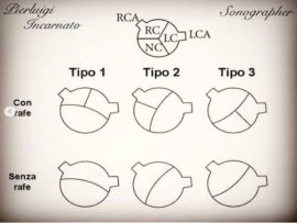
Bicuspid valve Type 2 without Rafe, with formation of bacterial endocarditis. Pierluigi Incarnato, TFCPC Cardiovascular and Abdominal Techinician Sonographer, Cardiac Physiologist at San Michele Nursing Home (Italy)
Read More
How do we know something is normal if we haven't seen the abnormality first? Normal MV vs a metallic MV replacement. The echo lady; Lorena De Vanna, is a..
Read More
Are most commonly seen between six and 10 days following an acute myocardial infarction (MI). They occur at the left ventricular apex and are more common following an anterior wall..
Read More
Aortic dissection is when the weakened wall of the aorta tears, causing blood to leak between the layers that makes up the wall. This can happen suddenly or slowly over..
Read More
A dilated aortic root is when the first section of the aorta, where the aortic valve resides, becomes enlarged. When this enlargement reaches a critical size, there is a risk..
Read More
Normal aortic valve opening properly Vs a heavily calcified aortic valve with severe stenosis. The echo lady; Lorena De Vanna, is a cardiac and respiratory physiologist graduated from the..
Read More
Left ventricular (LV) hypertrabeculation is defined by the presence of three or more trabeculations apically and up to the level of papillary muscles, seen in one echocardiographic view. It can..
Read More
The echo lady; Lorena De Vanna, is a cardiac and respiratory physiologist graduated from the Central University of Venezuela. She currently holds British Society of Echocardiography accreditation and works..
Read More
52 years old male patient with constrictive pericarditis after a CABG in 2018. Constrictive pericarditis is a potentially curable condition caused by a variety of situations which result in inflamed,..
Read More
Septal Occluder is a percutaneous, transcatheter, atrial septal defect closure device intended for the occlusion of atrial septal defects (ASD). ⠀ Patients indicated for ASD closure have echocardiographic evidence of..
Read More

Abstract An asymptomatic patient presented at our hospital exhibiting a Brugada electrocardiography pattern with coronary artery fistulas. Coronary artery fistula is a congenital or acquired rare abnormal condition with increased..
Read More
Abstract Objectives To investigate the prevalence and quantity of aortic valve calcium (AVC) in two large cohorts, stratified according to age and lipoprotein(a) (Lp(a)), and to assess the association between Lp(a)..
Read More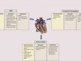
Abstract Myocardial fibrosis, seen in ischemic and nonischemic cardiomyopathies, is associated with adverse cardiac outcomes. Noninvasive imaging plays a key role in early identification and quantification of myocardial fibrosis with..
Read More
Abstract Despite multiple attempts to develop a unifying hypothesis that explains the pathophysiology of heart failure with a reduced ejection fraction (HFrEF), no single conceptual model has withstood the test..
Read More
Abstract Accurate echocardiographic evaluation of the systemic is challenging because of its specific morphology and contraction patterns. We present a detailed multimodality assessment of the systemic right ventricle, analyze the relative..
Read More
This program details the RESP and PRESERVE scores for predicting survival with the use of V-V ECMO in the ARDS patient. The program also details a single practice-multi, center program's..
Read More
The echo lady; Lorena De Vanna, is a cardiac and respiratory physiologist graduated from the Central University of Venezuela. She currently holds British Society..
Read More
The echo lady; Lorena De Vanna, is a cardiac and respiratory physiologist graduated from the Central University of Venezuela. She currently holds British Society..
Read More

