Transesophageal Echocardiography Guided Veno-Pulmonary Extracorporeal Membrane Oxygenation Cannula Insertion Technique
Abstract
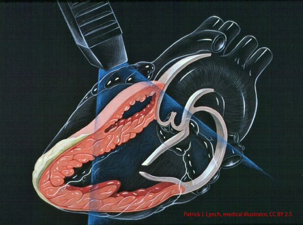
For single-lumen veno-pulmonary extracorporeal membrane oxygenation (VP ECMO) cannulation in percutaneous technique, vascular ultrasound (U/S) and transesophageal echocardiography (TEE) guidance were used to insert a 7 Fr pulmonary artery catheter (PAC) and an Amplatz Super Stiff (Boston Scientific) 0.035 inch × 260 cm guide wire (through the PAC) into the pulmonary artery (PA). The PAC was removed, and the Amplatz wire was kept in position. After vascular dilation, a long flexible 19 Fr 50 cm single-stage ECMO cannula was inserted over the stiff wire with its tip into the PA, and a 23 Fr 55 cm multistage drainage cannula into the right femoral vein with the tip terminating in the right atrium (RA). Transesophageal echocardiography was utilized for positioning and verification of cannula placement. We propose to use the following midesophageal (ME) views: “ME Bicaval” (Figure 1A), “ME RV inflow-outflow” (Figure 1B), and “ME asc aortic SAX” (Figure 1C) views. The PAC, Amplatz wire, and ECMO cannula may be viewed passing through the superior vena cava, the RA, the tricuspid valve, the RV, the right ventricular outflow tract, and through the pulmonic valve into the main PA. These three TEE views fully suffice for placement of the cannula. There are no recommended three-dimensional (3D) TEE views. We acquired the PA in short axis with the cannula tip visualized (Figure 1D), and in long axis view with the ECMO cannula traversing the pulmonic valve into the main PA (Figure 1E). These two-dimensional (2D) and 3D-TEE views provided real-time visualization which added safety and precision to cannula placement, but also verified the cannula position relative to its location within the PA and may also be useful in the assessment of the integrity of valvular cusps. A preoperative and postoperative chest radiograph (Figure 1F) and TEE exam may be useful for diagnostic purposes and to exclude potential postoperative complications, like wire perforation or pericardial effusion. In addition, early postoperative Chest radiograph (CXR) imaging should closely evaluate for unbalanced pulmonary arterial flow, and early detection of development of unilateral pulmonary edema. In our case, cannulation was successful and without complication, and enabled the patient to reach definitive treatment.