
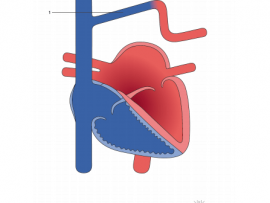

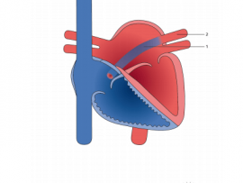
Partial anomalous pulmonary vein connection (PAPVC) is a rare congenital abnormal cardiac defect involving the pulmonary veins draining into the right atrium (RA) directly or indirectly by venous connection. Nowadays, these patients are diagnosed when still..
Read More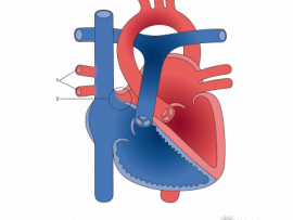
Partial anomalous venous connection (PAPVC) is a rare congenital heart disease where the blood flow from one or more pulmonary veins (but not all) returns to the right atrium or systemic venous circulation and is..
Read More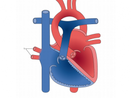
Partial anomalous pulmonary venous connection (PAPVC) is a rare congenital cardiac defect. As the name suggests, in PAPVC, the blood flow from a few of the pulmonary veins return to..
Read More
Partial anomalous pulmonary venous connection (PAPVC) is a rare congenital cardiac defect. As the name suggests, in PAPVC, the blood flow from a few of the pulmonary veins return to the right atrium instead..
Read More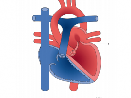
Barlow’s syndrome has become a regular, often-used and very often misused diagnosis. Its description followed extensive, prolonged and detailed clinical observation by JB Barlow and his co-workers. This major research..
Read More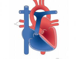
Partial anomalous venous connection (PAPVC) is a rare congenital heart disease where the blood flow from one or more pulmonary veins (but not all) returns to the right atrium or..
Read More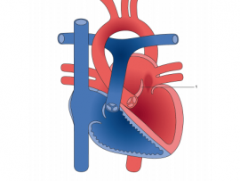
Mitral valve prolapse, also called MVP, is a condition in which the two valve flaps of the mitral valve don't close smoothly or evenly, but bulge (prolapse) upward into the..
Read More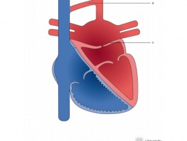
ECMO, El éxito de una terapia MÓDULO II – “ECMO, state of the art” MODERADOR Principal: Dr. Gregorio Rábago MD. Servicio Cirugía Cardiaca C.U.N., Pamplona/Madrid MODERADOR Chat: Dr. José Lopez...
Read More
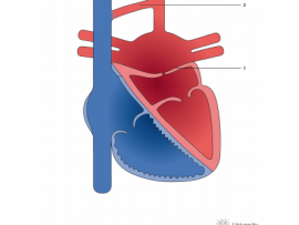

Cor triatriatum sinister is a relatively rare congenital condition in which the left atrium is bisected by a fibromuscular membrane into two distinct chambers. There are multiple hypotheses for the..
Read More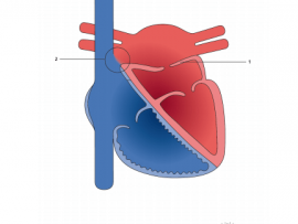
Cor Triatriatum Sinister is a rare congenital abnormality, usually diagnosed in childhood; few cases remain asymptomatic and are diagnosed in adulthood. In this review article we focus on classification and..
Read More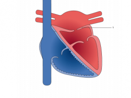
The cor triatriatum sinister is a rare congenital heart defect, the product of an embryonic defect in the incorporation of the common pulmonary vein to the left atrium, this being..
Read More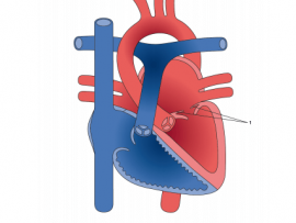
Supravalvar mitral ring is a circumferential ridge or membrane arising from the left atrial wall overlying the mitral valve and frequently attached to the mitral valve. Variable in thickness and extent,..
Read More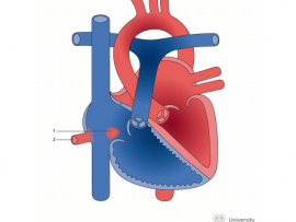
Background Sinus venosus defect (SVD) is an unusual type of interatrial communication (IAC) and is virtually always associated with partial anomalous pulmonary venous drainage (PAPVD) of the right pulmonary veins..
Read More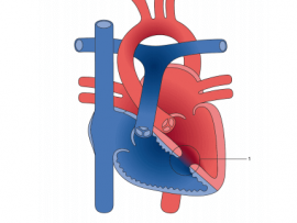
A ventricular septal defect (VSD) is a communication between the interventricular chambers. Muscular ventricular septal defects have exclusively muscular borders and are more likely to be multiple. Their location and..
Read More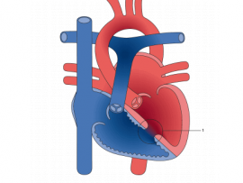
A ventricular septal defect (VSD) is an opening in the interventricular septum, causing a shunt between ventricles. Large defects result in a significant left-to-right shunt and cause dyspnea with feeding..
Read More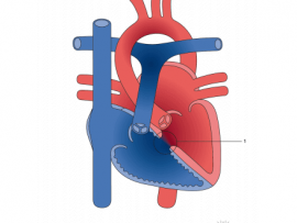
Perimembranous ventricular septal defects (VSDs) are located in the left ventricle outflow tract beneath the aortic valve. They are the most common subtype in the United States, occurring in 75-80% of..
Read More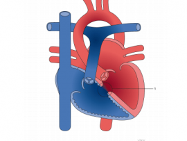
Perimembranous ventricular septal defects (VSDs) are located in the left ventricle outflow tract beneath the aortic valve. They are the most common VSD subtype in the United States, occurring in 75-80% of cases. Defects may extend into..
Read More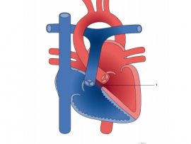
Doubly committed subarterial ventricular septal defect (VSD) is a unique type of VSD, located beneath the aortic and pulmonary valves, accounting for about 5–7% of all VSD autopsy findings. It is also called supracristal or infundibular VSD..
Read More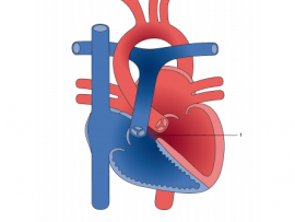
Background: The morphology of ventricular septal defects (VSDs) that are doubly committed and juxtaarterial places the patient at risk for aortic valvar prolapse and aortic valvar insufficiency (AI). Surgical repair of..
Read More
Atrioventricular septal defects (AVSD) are a relatively common family of congenital heart defects. Also known as atrioventricular canal defects or endocardial cushion defects, they account for about 5 percent of..
Read More
Female infant; born on term with 2.6 kg, normal delivery. Mother; fourth pregnancy, denied the use of medication during pregnancy. Presented respiratory discomfort at birth, evolving with neonatal infection and..
Read More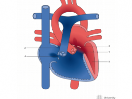
Background: Management of right-dominant atrioventricular septal defect (AVSD) remains a challenge given the spectrum of ventricular hypoplasia. The purpose of this study was to assess whether reported echocardiographic indices and additional..
Read More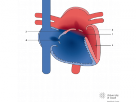
Unbalance in atrioventricular septal defect can be found in more than one anatomic level and in different degrees at each level. The definition of “unbalance” has historically been focused in..
Read More
Repair for tetralogy of Fallot (TOF) with complete atrioventricular septal defect (CAVSD) has been reported with good early and intermediate outcomes. Morbidity, however, remains significantly high. To date, repair of..
Read More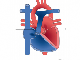
Atrioventricular (AV) septal defect consists of an ostium primum type atrial septal defect and a common AV valve, with or without an associated inlet (AV septal type) ventricular septal defect..
Read More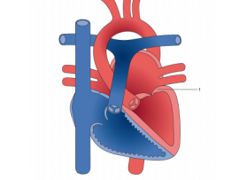
syndrome is a unique form of left-sided heart disease characterized by aortic outflow , dilated , dysplastic/incompetent mitral valve, and a restrictive/intact . Patients with this constellation of abnormalities have been managed in..
Read More