Pierluigi Incarnato, TFCPC Cardiovascular and Abdominal Techinician Sonographer, Cardiac Physiologist at San Michele Nursing Home (Italy)
Read More
Giant aorta from chronic aortic dissection: The true light and false trombised light is appreciated. Furthermore it is possible to observe the breach of entrance which feeds and supplies the..
Read More
Type 1R Type 4LR Pierluigi Incarnato, TFCPC Cardiovascular and Abdominal Techinician Sonographer, Cardiac Physiologist at San Michele Nursing Home (Italy)
Read More
Pierluigi Incarnato, TFCPC Cardiovascular and Abdominal Techinician Sonographer, Cardiac Physiologist at San Michele Nursing Home (Italy)
Read More
Pierluigi Incarnato, TFCPC Cardiovascular and Abdominal Techinician Sonographer, Cardiac Physiologist at San Michele Nursing Home (Italy)
Read More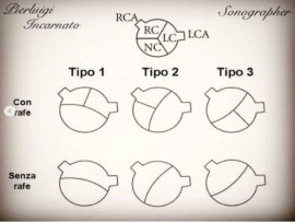
Bicuspid valve Type 2 without Rafe, with formation of bacterial endocarditis. Pierluigi Incarnato, TFCPC Cardiovascular and Abdominal Techinician Sonographer, Cardiac Physiologist at San Michele Nursing Home (Italy)
Read More
The takotsubo syndrome also called hypertrophic stress cardiomyopathy or shattered heart syndrome, is a clinical entity characterized by dysfunction of the left ventricle that manifests itself with symptoms that can..
Read More
Post-infarct patient with akinesia of the left ventricular apex, after about ten days from the first echocardium performed immediately after angioplasty, an important ecocontrast is evident in the left cavity..
Read More
Persistent left upper quarry in the long axis parasternal it is shown the dilated coronary sinus due to the persistence of the left superior vena cava, during the examination according..
Read More
Pierluigi Incarnato, TFCPC Cardiovascular and Abdominal Techinician Sonographer, Cardiac Physiologist at San Michele Nursing Home (Italy)
Read More
Pierluigi Incarnato, TFCPC Cardiovascular and Abdominal Techinician Sonographer, Cardiac Physiologist at San Michele Nursing Home (Italy)
Read More
Pierluigi Incarnato, TFCPC Cardiovascular and Abdominal Techinician Sonographer, Cardiac Physiologist at San Michele Nursing Home (Italy)
Read More
Pierluigi Incarnato, TFCPC Cardiovascular and Abdominal Techinician Sonographer, Cardiac Physiologist at San Michele Nursing Home (Italy)
Read More
Pierluigi Incarnato, TFCPC Cardiovascular and Abdominal Techinician Sonographer, Cardiac Physiologist at San Michele Nursing Home (Italy)
Read More
Pierluigi Incarnato, TFCPC Cardiovascular and Abdominal Techinician Sonographer, Cardiac Physiologist at San Michele Nursing Home (Italy)
Read More
Mohammed Zidan, MBBCH, M.Sc Cardiology (Cardiology, Echocardiography and interventional Cardilogy Specialist at Al-Azhar university), Cairo, Egypt.
Read More
Large cystic RA mass Mohammed Zidan, MBBCH, M.Sc Cardiology (Cardiology, Echocardiography and interventional Cardilogy Specialist at Al-Azhar university), Cairo, Egypt.
Read More
1. Modified 4chamver view due to all cardiac chamber dilation. 2. Huge LA due to sever mitral stenosis. 3. Sever rheumatic mitral stenosis with diastolic AML doaming. 4. Sever LA..
Read More
Severe Mitral Stenosis and Moderate Aortic Regurgitation Mohammed Zidan, MBBCH, M.Sc Cardiology (Cardiology, Echocardiography and interventional Cardilogy Specialist at Al-Azhar university), Cairo, Egypt.
Read More
TEE shows Hugely dilated Left Atrium as a consequence of Rhematic Severe Mitral Stenosis, spontaneous Echo Contrast Grade IV “pre thrombus formation “ Mohammed Zidan, MBBCH, M.Sc Cardiology (Cardiology,..
Read More
45 years old female patient with congestive Heart Failure “EF=22%” and history of stroke , the Echocardiography shows multiple big thrombi in the Heart chambers and moderate pericardial effusion ..
Read More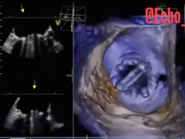
Severe Mitral Stenosis and Moderate Aortic Regurgitation Mohammed Zidan, MBBCH, M.Sc Cardiology (Cardiology, Echocardiography and interventional Cardilogy Specialist at Al-Azhar university), Cairo, Egypt.
Read More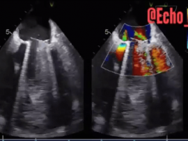
Paravalvular Leakage at 3 o’clock of Mitral Valve. Mohammed Zidan, MBBCH, M.Sc Cardiology (Cardiology, Echocardiography and interventional Cardilogy Specialist at Al-Azhar university), Cairo, Egypt.
Read More
Mohammed Zidan, MBBCH, M.Sc Cardiology (Cardiology, Echocardiography and interventional Cardilogy Specialist at Al-Azhar university), Cairo, Egypt.
Read More
72 years ald female patient admitted to hospital because of delirium and hypertensive crises. In TTE we found clearly suspicious finding on the RCC of the Aortic Valve. For further..
Read More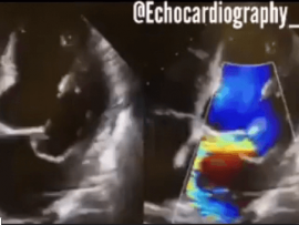
- Barlows prolapse. - Dilated left chambers, PMVL prolapse+ severe eccentric MR, Coanda effect, minimal pericardial effusion. - PML prolapse, severe MR eccentric jet, Dilated LA, Normal Lv systolic function.....
Read More
Mohammed Zidan, MBBCH, M.Sc Cardiology (Cardiology, Echocardiography and interventional Cardilogy Specialist at Al-Azhar university), Cairo, Egypt.
Read More
A 22-year-old man presented with palpitations and chest discomfort. Echocardiography revealed an enlarged, thickened mitral-valve leaflet with systolic prolapse of multiple segments (shown in a video), and a diagnosis of..
Read More