
In this video you can see patient with early post operative constrictive pericarditis and fungal mediastinal infection undergone thoracotomy and pericardiectomy as well as irrigation [gallery ids="29682,29686,29690"] Dr.Sam Zeraatian..
Weiterlesen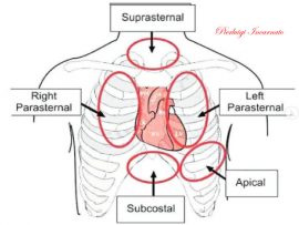
Pierluigi Incarnato, TFCPC Cardiovascular and Abdominal Techinician Sonographer, Cardiac Physiologist at San Michele Nursing Home (Italy)
Weiterlesen
Patient with Left atrial tumor. Dr.Sam Zeraatian Nejad Davani, CEO of rockingham medical research ADKWA center as well as Head of IUMS transplant and cardiovascular surgery department. ..
Weiterlesen
Aortic dissection is a serious condition in which there is a tear in the wall of the major artery carrying blood out of the heart (aorta). As the tear extends..
Weiterlesen
Apical thrombosis after previous acute myocardial infarction. Pierluigi Incarnato, TFCPC Cardiovascular and Abdominal Techinician Sonographer, Cardiac Physiologist at San Michele Nursing Home (Italy)
Weiterlesen
Nodule of Aranzio Anatomy of the aortic valve Pierluigi Incarnato, TFCPC Cardiovascular and Abdominal Techinician Sonographer, Cardiac Physiologist at San Michele Nursing Home (Italy)
Weiterlesen
Video Atrium Measurements Cardiovascular Sonographer Pierluigi Incarnato, TFCPC Cardiovascular and Abdominal Techinician Sonographer, Cardiac Physiologist at San Michele Nursing Home (Italy)
Weiterlesen
Pierluigi Incarnato, TFCPC Cardiovascular and Abdominal Techinician Sonographer, Cardiac Physiologist at San Michele Nursing Home (Italy)
Weiterlesen
[gallery ids="22031"] States that the volume of blood that passes through the mitral valve should be equal to the volume that passes through the aortic valve. SV = systolic range;..
Weiterlesen
In the last years, new trends on patient diagnosis for admission in cardiac intensive care unit (CICU) have been observed, shifting from acute myocardial infarction or acute heart failure to..
Weiterlesen
Apical akinesia in a patient undergoing primary PCI of residual IVA, with critical stenosis of CX and CTO of the right coronary. Pierluigi Incarnato, TFCPC Cardiovascular and Abdominal Techinician Sonographer,..
Weiterlesen
Severe mitral Stenosis insufficiency of the rheumatic type; doming of the flaps with hypomobility of the LMP. It can be seen Tunnel of the mitral inflow with minimal anatomical opening..
Weiterlesen
Background: McConnell's sign (right ventricular free wall [RV] hypokinesia with apical sparing on echocardiography) is often described as very specific for the diagnosis of pulmonary embolism (PE). In the video..
Weiterlesen
Learning points: The tricuspid valve is the largest of the heart valves . Its anatomical structure is complex and constituted of the 3 leaflets, the annulus, the chordae, the papillary..
Weiterlesen
AORTIC DISSECTION TYPE A‼️ All the didactics are on this fantastic video case and one of my previous post showing another type A dissection clinical case. From (twitter) Alex :..
Weiterlesen
Patient around 30yo, outpatient evaluated for heart txp. This is a congenital condition where the IVS fails to form correctly , and thus the blood from the atria is mixed..
Weiterlesen
this was a 28yo woman with this condition, where the aorta is still communicating with the pulmonary artery. From LopezOpitz (twitter) Alex : is a last year medical student,..
Weiterlesen
Intramyocardial dissection is a rare complication of myocardial infarction, trauma, and percutaneous intervention. It is usually caused by hemorrhagic dissection among the spiral myocardial fibers. This is the case of..
Weiterlesen
This is how a normal aortic bioprosthetic valve is showed on echo. This type of valve (which doesn’t need an INR between 2-3) is generally used in old people (with..
Weiterlesen
Amyloid cardiomyopathy is a restrictive infiltrative disease that results in severe LV diastolic dysfunction and moderate LV systolic dysfunction. Classically, one may see a significant biatrial enlargement along with leaky..
Weiterlesen
ARVD, sometimes also called ARVC (C for Cardiomyopathy), is a rare condition where fibrofatty collagen tissue replaces the normal RV wall. Less commonly, this process involves the LV. Classically the..
Weiterlesen
In LVNC, the lower left chamber of the heart, called the left ventricle, contains bundles or pieces of muscle that extend into the chamber. These pieces of muscles are called..
Weiterlesen
It can be asymptomatic (commonly diagnosed as an incidental finding on an echocardiogram) or cause symptoms and embolize (same as atrial myxoma, the most frequent benign tumor). Characteristically, it is..
Weiterlesen
This is a condition where pulmonary etiology (such as pulmonary hypertension, PHT) causes progressive right heart failure (dilation and/or hypertrophy) only, with unaffected left heart. It can be derived from..
Weiterlesen
A 50-year-old patient presents following a motor vehicle accident. An echocardiogram is ordered to evaluate for shortness of breath. What is indicated by the arrow in the image? The..
Weiterlesen
Subvalvular aortic stenosis (SAS) is one of the common adult congenital heart diseases, with a prevalence of 6.5%. It is usually diagnosed in the first decade of life. Echocardiography is..
Weiterlesen
Learning points: Hydatidosis or cystic echinococcosis is caused by infection with the metacestode stage of the tapeworm Echinococcus (family Taeniidae). The adult tapeworm is usually found in dogs or other..
Weiterlesen
How to suspect pulmonary vascular disease ?! Here are some useful points: - Estimation of systolic pulmonary pressure - Pulmonary artery size and doppler - Right atrium / ventricle and..
Weiterlesen
Pierluigi Incarnato, TFCPC Cardiovascular and Abdominal Techinician Sonographer, Cardiac Physiologist at San Michele Nursing Home (Italy)
Weiterlesen
Pierluigi Incarnato, TFCPC Cardiovascular and Abdominal Techinician Sonographer, Cardiac Physiologist at San Michele Nursing Home (Italy)
Weiterlesen