Epicardial Fat – Case report
- Zuhause
- Echocardiography
- Aktuelle Seite
A 50-year-old patient presents following a motor vehicle accident. An echocardiogram is ordered to evaluate for shortness of breath. What is indicated by the arrow in the image?
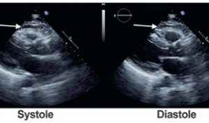
The arrow indicates a space anterior to the right ventricle, which is of mixed echogenicity. This is a typical finding in individuals with epicardial fat. Epicardial fat is generally seen in between the right ventricular wall and chest wall. It is an area that is almost echo-free and moves with the right ventricle. CT scan of the same patient clearly delineates this structure as epicardial fat due to its low Hounsfield units (dark appearance). A pericardial effusion is usually echo-free, although a pericardial hematoma can have mixed echogenicity
Alex : is a last year medical student, very passionate about echo, POCUS and hand-held ultrasound devices.
He posts YouTube reviews of these, as well as interesting cases in Instagram (@mastersofultrasound) and Twitter (@mastersofultrasound).
Wir verwenden Cookies, um Ihnen die bestmögliche Benutzererfahrung zu bieten. Durch die weitere Nutzung unserer Website stimmen Sie deren Nutzung zu. Mehr erfahren