
Patient around 30yo, outpatient evaluated for heart txp. This is a congenital condition where the IVS fails to form correctly , and thus the blood from the atria is mixed..
Read More
this was a 28yo woman with this condition, where the aorta is still communicating with the pulmonary artery. From LopezOpitz (twitter) Alex : is a last year medical student,..
Read More
Intramyocardial dissection is a rare complication of myocardial infarction, trauma, and percutaneous intervention. It is usually caused by hemorrhagic dissection among the spiral myocardial fibers. This is the case of..
Read More
Cor Triatriatum occurs when either the right or left atrium has a flap or tissue or membrane that divides it into two chambers. The classical "Cor Triatrium sinister" is shown..
Read More
This is how a normal aortic bioprosthetic valve is showed on echo. This type of valve (which doesn’t need an INR between 2-3) is generally used in old people (with..
Read More
Amyloid cardiomyopathy is a restrictive infiltrative disease that results in severe LV diastolic dysfunction and moderate LV systolic dysfunction. Classically, one may see a significant biatrial enlargement along with leaky..
Read More
ARVD, sometimes also called ARVC (C for Cardiomyopathy), is a rare condition where fibrofatty collagen tissue replaces the normal RV wall. Less commonly, this process involves the LV. Classically the..
Read More
In LVNC, the lower left chamber of the heart, called the left ventricle, contains bundles or pieces of muscle that extend into the chamber. These pieces of muscles are called..
Read More
It can be asymptomatic (commonly diagnosed as an incidental finding on an echocardiogram) or cause symptoms and embolize (same as atrial myxoma, the most frequent benign tumor). Characteristically, it is..
Read More
Here you can see an amazing case of double valve endocarditis, involving the tricuspid and mitral valves. (apical 4 chambers) displays both affected valves focuses on the..
Read More
This is a condition where pulmonary etiology (such as pulmonary hypertension, PHT) causes progressive right heart failure (dilation and/or hypertrophy) only, with unaffected left heart. It can be derived from..
Read More
A 50-year-old patient presents following a motor vehicle accident. An echocardiogram is ordered to evaluate for shortness of breath. What is indicated by the arrow in the image? The..
Read More
Relacion pulmonar-sistemica, mejor conocida como la relacion Qp/Qs. - Video de como medir el TSVI aqui: - Video de como medir el volumen eyectado aqui: Pulmonary-systemic flow ratio, better..
Read More
Alex : is a last year medical student, very passionate about echo, POCUS and hand-held ultrasound devices. He posts YouTube reviews of these, as well as interesting..
Read More
Comprehensive transthoracic Echocardiography examination protocol. Windows, Imaging planes and views
Alex : is a last year medical student, very passionate about echo, POCUS and hand-held ultrasound devices. He posts YouTube reviews of these, as well as interesting cases..
Read More
TEE is a semi-invasive technique used to assess left heart cavities with better resolution and angles than conventional TTE, specially when studying valvulopathies, interventricular communications or atrial masses. It is..
Read More
Subvalvular aortic stenosis (SAS) is one of the common adult congenital heart diseases, with a prevalence of 6.5%. It is usually diagnosed in the first decade of life. Echocardiography is..
Read More
Learning points: Hydatidosis or cystic echinococcosis is caused by infection with the metacestode stage of the tapeworm Echinococcus (family Taeniidae). The adult tapeworm is usually found in dogs or other..
Read More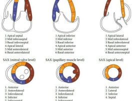

En este video estaré dándoles mi opinión acerca del dispositivo de ultrasonido KOSMOS. Este no es un video patrocinado y las opiniones emitidas son personales. In this video I..
Read More
Endocarditis related to PM-lead infection is a rare but serious complication of permanent transvenous pacing. The reported incidence after permanent endocardial PM implantation varies in the literature from 0.13% to..
Read More
Carol Mitchell, PhD, RDMS, RDCS, RVT, RT(R), ACS, FASE, reviews Guidelines for Performing a Comprehensive Transthoracic Echocardiographic Examination in Adults: Recommendations from the American Society of Echocardiography. Peter Rahko, MD,..
Read More
Rebecca T. Hahn, MD, FASE reviews Guidelines for Performing a Comprehensive Transesophageal Echocardiographic Examination: Recommendations from ASE and SCA
Read More
42 years old male patient with hypertrophic cardiomyopathy. - Septum (2.9cm). - Posterior wall (2.7cm). What is HCM? In HCM the muscular walls of the heart’s ventricles become thickened. HCM..
Read More
Abstract A systematic approach to transoesophageal echocardiography (TOE) is essential to ensure that no pathology is missed during a study. In addition, a standardised approach facilitates the education and..
Read More
Abstract There have been significant advances in the field of echocardiography with the introduction of a number of new techniques into standard clinical practice. Consequently, a ‘standard’ echocardiographic examination has..
Read More
En este video voy a hablar del gasto cardiaco y como obtenerlo por medio del ecocardiograma. In this video I am going to talk about the cardiac output and how..
Read More
Takotsubo cardiomyopathy, also known as stress cardiomyopathy and "broken heart syndrome," is a sudden, transient cardiac syndrome that involves dramatic left ventricular apical akinesis and mimics acute coronary syndrome (ACS). It was..
Read More
Lipomatous hypertrophy of the interatrial septum (LHIS) is a benign cardiac mass characterised as a non-encapsulated mass of fatty tissue that infiltrates the atrial septum. ⠀ Although once described as..
Read More
Spontaneous echocardiographic contrast (SEC): is a phenomenon of discrete reflections appearing in the blood inside the cardiac chambers, cavities or vessels without previous injection of echocontrast media or fluids containing..
Read More