Tricuspid atresia with patent foramen ovale (PFO)
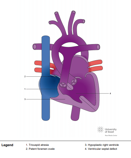
The normal pumping chambers of the heart (the ventricles) must have an inflow valve to let blood in, a good-sized pumping chamber and an outflow to allow blood to exit the chamber during contraction. In tricuspid atresia, the tricuspid valve, which lies between the heart’s upper right chamber (the right atrium) and lower right chamber (the right ventricle), does not develop. As a result, it prevents oxygen-depleted blood that is returning to the right atrium from the body from directly flowing into the right ventricle (the chamber that normally pumps blood to the lungs where it picks up oxygen). Instead, the venous blue blood must pass through a hole between the upper filling chambers (the atria), where it then mixes with oxygen-rich blood returning from the lungs via the pulmonary veins. This combined blood then passes to the lower left pumping chamber.
Commonly in tricuspid atresia, the right ventricle may be small because blood flow to the right ventricle is blocked by the non-existent tricuspid valve, and the lower right chamber does not receive a normal amount of blood. In many situations, there is a hole between the two lower pumping chambers (a ventricular septal defect, or VSD). Some of the blood ejected by the left ventricle passes out to the aorta on its way to the body. Some of the blood may cross the ventricular septal defect to go to the lungs. Additionally, there may be muscular narrowing in the right ventricle under the pulmonary valve that keeps blood from entering the pulmonary arteries. In some situations, the main blood vessels exiting the heart (the aorta and pulmonary arteries) may be connected to the wrong pumping chamber. This is called a transposition.